6L30

Crystal structure of the epithelial cell transforming 2 (ECT2)


Experimental Data Snapshot

  • Method: X-RAY DIFFRACTION
  • Resolution: 2.80 Å
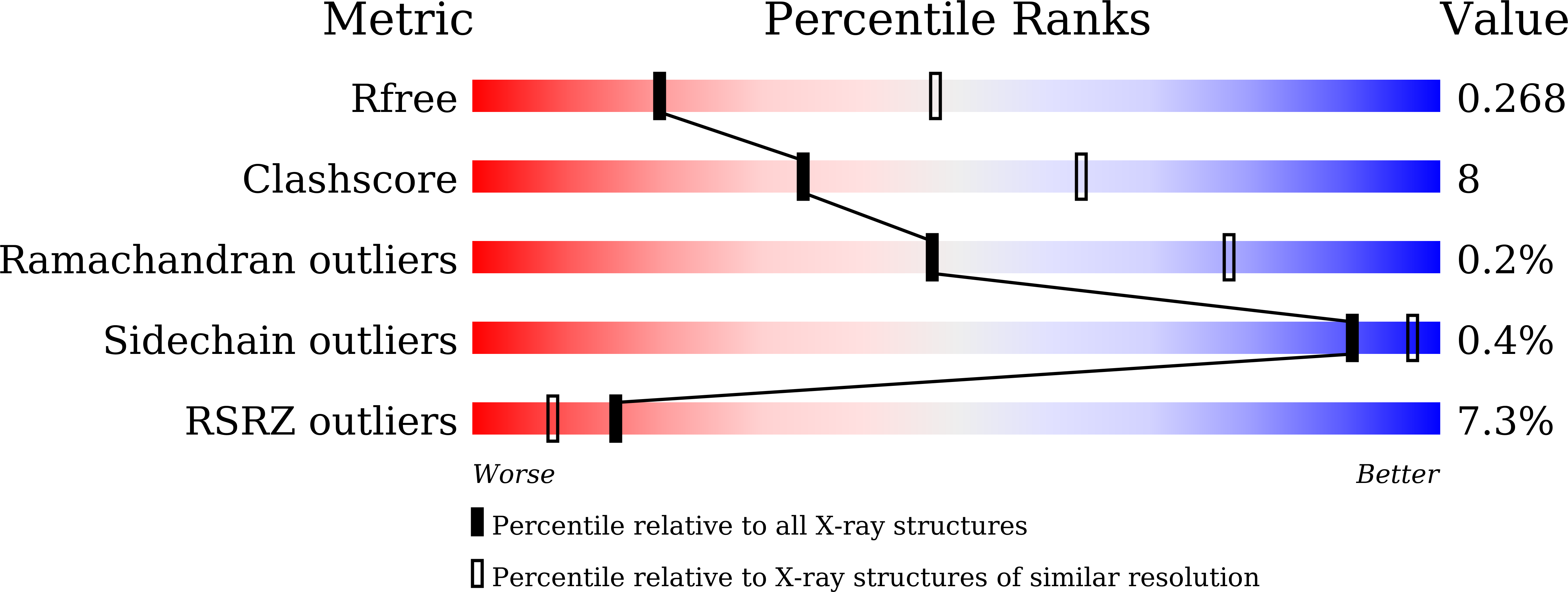
  • R-Value Free: 0.269 
  • R-Value Work: 0.237 
  • R-Value Observed: 0.238 

wwPDB Validation   3D Report Full Report


This is version 1.3 of the entry. See complete history


Literature

Structure and regulation of human epithelial cell transforming 2 protein.

Chen, M.Pan, H.Sun, L.Shi, P.Zhang, Y.Li, L.Huang, Y.Chen, J.Jiang, P.Fang, X.Wu, C.Chen, Z.

(2020) Proc Natl Acad Sci U S A 117: 1027-1035

  • DOI: https://doi.org/10.1073/pnas.1913054117
  • Primary Citation of Related Structures:  
    6L30

  • PubMed Abstract: 

    Epithelial cell transforming 2 (Ect2) protein activates Rho GTPases and controls cytokinesis and many other cellular processes. Dysregulation of Ect2 is associated with various cancers. Here, we report the crystal structure of human Ect2 and complementary mechanistic analyses. The data show the C-terminal PH domain of Ect2 folds back and blocks the canonical RhoA-binding site at the catalytic center of the DH domain, providing a mechanism of Ect2 autoinhibition. Ect2 is activated by binding of GTP-bound RhoA to the PH domain, which suggests an allosteric mechanism of Ect2 activation and a positive-feedback loop reinforcing RhoA signaling. This bimodal RhoA binding of Ect2 is unusual and was confirmed with Förster resonance energy transfer (FRET) and hydrogen-deuterium exchange mass spectrometry (HDX-MS) analyses. Several recurrent cancer-associated mutations map to the catalytic and regulatory interfaces, and dysregulate Ect2 in vitro and in vivo. Together, our findings provide mechanistic insights into Ect2 regulation in normal cells and under disease conditions.


  • Organizational Affiliation

    Ministry of Education Key Laboratory of Protein Science, Tsinghua University, Beijing 100084, China.


Macromolecules
Find similar proteins by:  (by identity cutoff)  |  3D Structure
Entity ID: 1
MoleculeChains Sequence LengthOrganismDetailsImage
Protein ECT2707Homo sapiensMutation(s): 0 
Gene Names: ECT2
UniProt & NIH Common Fund Data Resources
Find proteins for Q9H8V3 (Homo sapiens)
Explore Q9H8V3 
Go to UniProtKB:  Q9H8V3
PHAROS:  Q9H8V3
GTEx:  ENSG00000114346 
Entity Groups  
Sequence Clusters30% Identity50% Identity70% Identity90% Identity95% Identity100% Identity
UniProt GroupQ9H8V3
Sequence Annotations
Expand
  • Reference Sequence
Experimental Data & Validation

Experimental Data

  • Method: X-RAY DIFFRACTION
  • Resolution: 2.80 Å
  • R-Value Free: 0.269 
  • R-Value Work: 0.237 
  • R-Value Observed: 0.238 
  • Space Group: I 2 2 2
Unit Cell:
Length ( Å )Angle ( ˚ )
a = 106.959α = 90
b = 130.579β = 90
c = 131.773γ = 90
Software Package:
Software NamePurpose
PHENIXrefinement
PDB_EXTRACTdata extraction
HKL-2000data reduction
HKL-2000data scaling
SHARPphasing

Structure Validation

View Full Validation Report


Entry History & Funding Information

Deposition Data


Funding OrganizationLocationGrant Number
National Natural Science Foundation of ChinaChina--

Revision History  (Full details and data files)

  • Version 1.0: 2020-01-01
    Type: Initial release
  • Version 1.1: 2020-01-15
    Changes: Database references
  • Version 1.2: 2020-01-29
    Changes: Database references
  • Version 1.3: 2023-11-22
    Changes: Data collection, Database references, Refinement description