6KFQ

Crystal structure of thermophilic rhodopsin from Rubrobacter xylanophilus


Experimental Data Snapshot

  • Method: X-RAY DIFFRACTION
  • Resolution: 1.84 Å
  • R-Value Free: 0.227 
  • R-Value Work: 0.193 
  • R-Value Observed: 0.194 

wwPDB Validation   3D Report Full Report


Ligand Structure Quality Assessment 


This is version 1.1 of the entry. See complete history


Literature

How Does a Microbial Rhodopsin RxR Realize Its Exceptionally High Thermostability with the Proton-Pumping Function Being Retained?

Hayashi, T.Yasuda, S.Suzuki, K.Akiyama, T.Kanehara, K.Kojima, K.Tanabe, M.Kato, R.Senda, T.Sudo, Y.Murata, T.Kinoshita, M.

(2020) J Phys Chem B 124: 990-1000

  • DOI: https://doi.org/10.1021/acs.jpcb.9b10700
  • Primary Citation of Related Structures:  
    6KFQ

  • PubMed Abstract: 

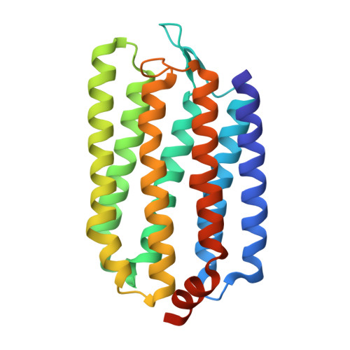
    We often encounter a case where two proteins, whose amino-acid sequences are similar, are quite different with regard to the thermostability. As a striking example, we consider the two seven-transmembrane proteins: recently discovered Rubrobacter xylanophilus rhodopsin (RxR) and long-known bacteriorhodopsin from Halobacterium salinarum (HsBR). They commonly function as a light-driven proton pump across the membrane. Though their sequence similarity and identity are ∼71 and ∼45%, respectively, RxR is much more thermostable than HsBR. In this study, we solve the three-dimensional structure of RxR using X-ray crystallography and find that the backbone structures of RxR and HsBR are surprisingly similar to each other: The root-mean-square deviation for the two structures calculated using the backbone C α atoms of the seven helices is only 0.86 Å, which makes the large stability difference more puzzling. We calculate the thermostability measure and its energetic and entropic components for RxR and HsBR using our recently developed statistical-mechanical theory. The same type of calculation is independently performed for the portions playing essential roles in the proton-pumping function, helices 3 and 7, and their structural properties are related to the probable roles of water molecules in the proton-transporting mechanism. We succeed in elucidating how RxR realizes its exceptionally high stability with the original function being retained. This study provides an important first step toward the establishment of a method correlating microscopic, geometric characteristics of a protein with its thermodynamic properties and enhancing the thermostability through amino-acid mutations without vitiating the original function.


  • Organizational Affiliation

    Institute of Advanced Energy , Kyoto University , Gokasho, Uji, Kyoto 611-0011 , Japan.


Macromolecules
Find similar proteins by:  (by identity cutoff)  |  3D Structure
Entity ID: 1
MoleculeChains Sequence LengthOrganismDetailsImage
Rhodopsin239Rubrobacter xylanophilus DSM 9941Mutation(s): 0 
Gene Names: Rxyl_2037
Membrane Entity: Yes 
UniProt
Find proteins for Q1AUE6 (Rubrobacter xylanophilus (strain DSM 9941 / NBRC 16129 / PRD-1))
Explore Q1AUE6 
Go to UniProtKB:  Q1AUE6
Entity Groups  
Sequence Clusters30% Identity50% Identity70% Identity90% Identity95% Identity100% Identity
UniProt GroupQ1AUE6
Sequence Annotations
Expand
  • Reference Sequence
Small Molecules
Ligands 3 Unique
IDChains Name / Formula / InChI Key2D Diagram3D Interactions
MPG (Subject of Investigation/LOI)
Query on MPG

Download Ideal Coordinates CCD File 
B [auth A]
C [auth A]
D [auth A]
E [auth A]
F [auth A]
B [auth A],
C [auth A],
D [auth A],
E [auth A],
F [auth A],
G [auth A],
H [auth A],
I [auth A],
J [auth A],
K [auth A],
L [auth A],
M [auth A],
N [auth A],
O [auth A],
P [auth A],
Q [auth A]
[(Z)-octadec-9-enyl] (2R)-2,3-bis(oxidanyl)propanoate
C21 H40 O4
JPJYKWFFJCWMPK-GDCKJWNLSA-N
RET (Subject of Investigation/LOI)
Query on RET

Download Ideal Coordinates CCD File 
R [auth A]RETINAL
C20 H28 O
NCYCYZXNIZJOKI-OVSJKPMPSA-N
SO4 (Subject of Investigation/LOI)
Query on SO4

Download Ideal Coordinates CCD File 
S [auth A],
T [auth A]
SULFATE ION
O4 S
QAOWNCQODCNURD-UHFFFAOYSA-L
Experimental Data & Validation

Experimental Data

  • Method: X-RAY DIFFRACTION
  • Resolution: 1.84 Å
  • R-Value Free: 0.227 
  • R-Value Work: 0.193 
  • R-Value Observed: 0.194 
  • Space Group: P 21 21 21
Unit Cell:
Length ( Å )Angle ( ˚ )
a = 29.93α = 90
b = 68.41β = 90
c = 107.91γ = 90
Software Package:
Software NamePurpose
PHENIXrefinement
XDSdata reduction
Cootmodel building
PHASERphasing

Structure Validation

View Full Validation Report


Ligand Structure Quality Assessment 


Entry History 

Deposition Data

Revision History  (Full details and data files)

  • Version 1.0: 2020-04-08
    Type: Initial release
  • Version 1.1: 2023-11-22
    Changes: Data collection, Database references, Refinement description