Protein surface charge effect on 3D domain swapping in cells for c-type cytochromes.
Yang, H., Yamanaka, M., Nagao, S., Yasuhara, K., Shibata, N., Higuchi, Y., Hirota, S.(2019) Biochim Biophys Acta Proteins Proteom 1867: 140265-140265
- PubMed: 31437585
- DOI: https://doi.org/10.1016/j.bbapap.2019.140265
- Primary Citation of Related Structures:
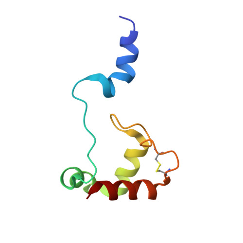
6K7C - PubMed Abstract:
Many c-type cytochromes (cyts) can form domain-swapped oligomers. The positively charged Hydrogenobacter thermophilus (HT) cytochrome (cyt) c 552 forms domain-swapped oligomers during expression in the Escherichia coli (E. coli) expression system, but the factors influencing the oligomerization remain unrevealed. Here, we found that the dimer of the negatively charged Shewanella violacea (SV) cyt c 5 exhibits a domain-swapped structure, in which the N-terminal helix is exchanged between protomers, similar to the structures of the HT cyt c 552 and Pseudomonas aeruginosa (PA) cyt c 551 domain-swapped dimers. Positively charged horse cyt c and HT cyt c 552 domain swapped during expression in E. coli, whereas negatively charged PA cyt c 551 and SV cyt c 5 did not. Oligomers were formed during expression in E. coli for HT cyt c 552 attached to either a co- or post-translational signal peptide for transportation through the cytoplasm membrane, but not for PA cyt c 551 attached to either signal peptide. HT cyt c 552 formed oligomers in E. coli in the presence and absence of rare codons. More oligomers were obtained from the in vitro folding of horse cyt c and HT cyt c 552 by the addition of negatively charged liposomes during folding, whereas the amount of oligomers for the in vitro folding of PA cyt c 551 and SV cyt c 5 did not change significantly by the addition. These results indicate that the protein surface charge affects the oligomerization of c-type cyts in cells; positively charged c-type cyts assemble on a negatively charged membrane, inducing formation of domain-swapped oligomers during folding.
Organizational Affiliation:
Division of Materials Science, Graduate School of Science and Technology, Nara Institute of Science and Technology, 8916-5 Takayama, Ikoma, Nara 630-0192, Japan.