6K1R

Crystal structure of Ketopantoate reductase from Pseudomonas aeruginosa in complex with NAD+ and ketopantoate


Experimental Data Snapshot

  • Method: X-RAY DIFFRACTION
  • Resolution: 2.55 Å
  • R-Value Free: 0.269 
  • R-Value Work: 0.244 
  • R-Value Observed: 0.245 

wwPDB Validation   3D Report Full Report


Ligand Structure Quality Assessment 


This is version 1.2 of the entry. See complete history


Literature

Crystallographic and biophysical analyses of Pseudomonas aeruginosa ketopantoate reductase: Implications of ligand induced conformational changes in cofactor recognition.

Choudhury, A.Khanppnavar, B.Datta, S.

(2021) Biochimie 

  • DOI: https://doi.org/10.1016/j.biochi.2021.10.015
  • Primary Citation of Related Structures:  
    6K1R

  • PubMed Abstract: 

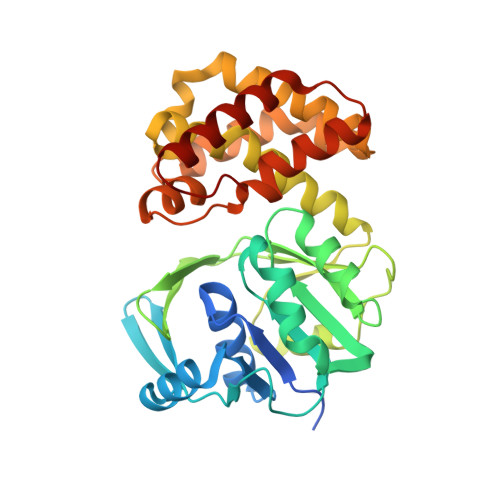
    Ketopantoate reductases (KPRs) catalyse NADPH-dependent reduction of ketopantoate to pantoate, the rate-limiting step of pantothenate biosynthetic pathway. In our recent study, we showed KPRs are under dynamic evolutionary selection and highlighted the possible role of ordered substrate binding kinetics for cofactor selection. To further delineate this at molecular level, here, we perform X-ray crystallographic and biophysical analyses of KPR in presence of non-canonical cofactor NAD + . In our structure, NAD + was found to be highly dynamic in catalytic pocket of KPR, which could attain stable conformation only in presence of ketopantoate. Further, isothermal calorimetric (ITC) titrations showed that affinity of KPR for ketopantoate is higher in presence of NADP + than in presence of NAD + and lowest in absence of redox cofactors. In sum, our results clearly depict two modes of redox cofactor selections in KPRs, firstly by specific salt bridge interactions with unique phosphate moiety of NADP + and secondly via ordered sequential heterotrophic cooperative binding of substrate ketopantoate.


  • Organizational Affiliation

    Department of Structural Biology and Bioinformatics, CSIR-Indian Institute of Chemical Biology (CSIR-IICB), Kolkata, India; Academy of Scientific and Innovative Research (AcSIR), India.


Macromolecules
Find similar proteins by:  (by identity cutoff)  |  3D Structure
Entity ID: 1
MoleculeChains Sequence LengthOrganismDetailsImage
Probable 2-dehydropantoate 2-reductase
A, B, C
323Pseudomonas aeruginosa PAO1Mutation(s): 0 
Gene Names: panE
EC: 1.1.1.169
UniProt
Find proteins for Q9HW09 (Pseudomonas aeruginosa (strain ATCC 15692 / DSM 22644 / CIP 104116 / JCM 14847 / LMG 12228 / 1C / PRS 101 / PAO1))
Explore Q9HW09 
Go to UniProtKB:  Q9HW09
Entity Groups  
Sequence Clusters30% Identity50% Identity70% Identity90% Identity95% Identity100% Identity
UniProt GroupQ9HW09
Sequence Annotations
Expand
  • Reference Sequence
Experimental Data & Validation

Experimental Data

  • Method: X-RAY DIFFRACTION
  • Resolution: 2.55 Å
  • R-Value Free: 0.269 
  • R-Value Work: 0.244 
  • R-Value Observed: 0.245 
  • Space Group: P 42 21 2
Unit Cell:
Length ( Å )Angle ( ˚ )
a = 129.995α = 90
b = 129.995β = 90
c = 155.105γ = 90
Software Package:
Software NamePurpose
PHENIXrefinement
PROTEUM PLUSdata reduction
PROTEUM PLUSdata scaling
PHASERphasing

Structure Validation

View Full Validation Report


Ligand Structure Quality Assessment 


Entry History & Funding Information

Deposition Data


Funding OrganizationLocationGrant Number
Department of Science & Technology (India)IndiaSB/SO/BB - 36/2014

Revision History  (Full details and data files)

  • Version 1.0: 2020-05-13
    Type: Initial release
  • Version 1.1: 2021-11-24
    Changes: Database references
  • Version 1.2: 2023-11-22
    Changes: Data collection, Refinement description