


Crystal structure of Human G6PD Canton
Au, S.W.N.To be published.
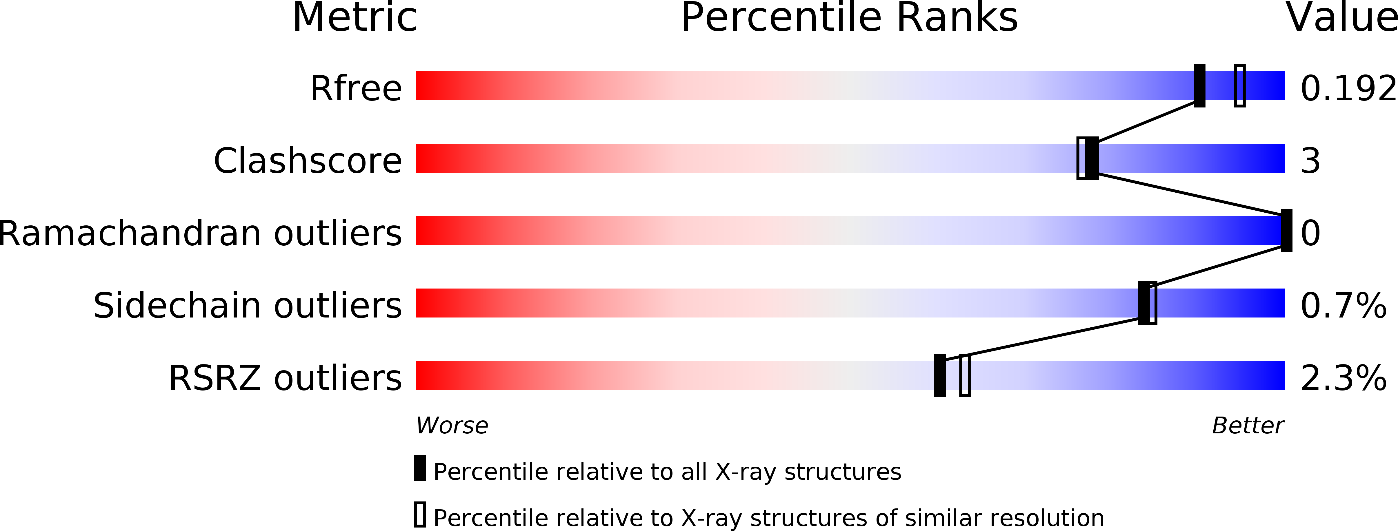
Experimental Data Snapshot
Entity ID: 1 | |||||
|---|---|---|---|---|---|
| Molecule | Chains | Sequence Length | Organism | Details | Image |
| Glucose-6-phosphate 1-dehydrogenase | 485 | Homo sapiens | Mutation(s): 1 Gene Names: G6PD EC: 1.1.1.49 | 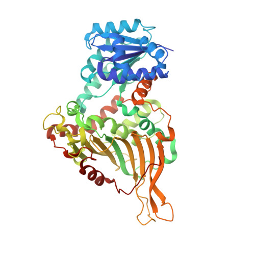 | |
UniProt & NIH Common Fund Data Resources | |||||
Find proteins for P11413 (Homo sapiens) Explore P11413 Go to UniProtKB: P11413 | |||||
PHAROS: P11413 GTEx: ENSG00000160211 | |||||
Entity Groups | |||||
| Sequence Clusters | 30% Identity50% Identity70% Identity90% Identity95% Identity100% Identity | ||||
| UniProt Group | P11413 | ||||
Sequence AnnotationsExpand | |||||
| |||||
| Ligands 1 Unique | |||||
|---|---|---|---|---|---|
| ID | Chains | Name / Formula / InChI Key | 2D Diagram | 3D Interactions | |
| NAP Query on NAP | B [auth A] | NADP NICOTINAMIDE-ADENINE-DINUCLEOTIDE PHOSPHATE C21 H28 N7 O17 P3 XJLXINKUBYWONI-NNYOXOHSSA-N |  | ||
| Length ( Å ) | Angle ( ˚ ) |
|---|---|
| a = 60.422 | α = 90 |
| b = 172.83 | β = 90 |
| c = 215.522 | γ = 90 |
| Software Name | Purpose |
|---|---|
| PHENIX | refinement |
| HKL-2000 | data reduction |
| SCALEPACK | data scaling |
| PHENIX | phasing |
RCSB PDB (citation) is hosted by
RCSB PDB is a member of the

