6JHE

Crystal Structure of Bacillus subtilis SigW domain 4 in complexed with -35 element DNA


Experimental Data Snapshot

  • Method: X-RAY DIFFRACTION
  • Resolution: 3.10 Å
  • R-Value Free: 0.289 
  • R-Value Work: 0.247 
  • R-Value Observed: 0.250 

wwPDB Validation   3D Report Full Report


This is version 1.1 of the entry. See complete history


Literature

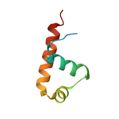
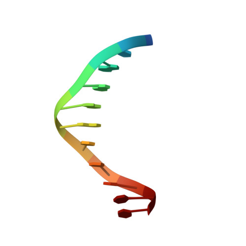
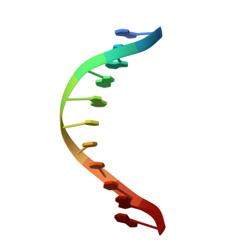
Structural analysis of the recognition of the -35 promoter element by SigW from Bacillus subtilis.

Kwon, E.Devkota, S.R.Pathak, D.Dahal, P.Kim, D.Y.

(2019) PLoS One 14: e0221666-e0221666

  • DOI: https://doi.org/10.1371/journal.pone.0221666
  • Primary Citation of Related Structures:  
    6JHE

  • PubMed Abstract: 

    Sigma factors are key proteins that mediate the recruitment of RNA polymerase to the promoter regions of genes, for the initiation of bacterial transcription. Multiple sigma factors in a bacterium selectively recognize their cognate promoter sequences, thereby inducing the expression of their own regulons. In this paper, we report the crystal structure of the σ4 domain of Bacillus subtilis SigW bound to the -35 promoter element. Purine-specific hydrogen bonds of the -35 promoter element with the recognition helix α9 of the σ4 domain occurs at three nucleotides of the consensus sequence (G-35, A-34, and G'-31 in G-35A-34A-33A-32C-31C-30T-29). The hydrogen bonds of the backbone with the α7 and α8 of the σ4 domain occurs at G'-30. These results elucidate the structural basis of the selective recognition of the promoter by SigW. In addition, comparison of SigW structures complexed with the -35 promoter element or with anti-sigma RsiW reveals that DNA recognition and anti-sigma factor binding of SigW are mutually exclusive.


  • Organizational Affiliation

    College of Pharmacy, Yeungnam University, Gyeongsan, Gyeongbuk, South Korea.


Macromolecules

Find similar proteins by:  (by identity cutoff)  |  3D Structure
Entity ID: 1
MoleculeChains Sequence LengthOrganismDetailsImage
ECF RNA polymerase sigma factor SigW64Bacillus subtilis subsp. subtilis str. 168Mutation(s): 0 
Gene Names: sigWybbLBSU01730
UniProt
Find proteins for Q45585 (Bacillus subtilis (strain 168))
Explore Q45585 
Go to UniProtKB:  Q45585
Entity Groups  
Sequence Clusters30% Identity50% Identity70% Identity90% Identity95% Identity100% Identity
UniProt GroupQ45585
Sequence Annotations
Expand
  • Reference Sequence

Find similar nucleic acids by:  Sequence   |   3D Structure  

Entity ID: 2
MoleculeChains LengthOrganismImage
DNA (5'-D(P*TP*TP*GP*AP*AP*AP*CP*CP*TP*TP*T)-3')11synthetic construct
Sequence Annotations
Expand
  • Reference Sequence

Find similar nucleic acids by:  Sequence   |   3D Structure  

Entity ID: 3
MoleculeChains LengthOrganismImage
DNA (5'-D(*AP*AP*AP*GP*GP*TP*TP*TP*CP*AP*A)-3')11synthetic construct
Sequence Annotations
Expand
  • Reference Sequence
Experimental Data & Validation

Experimental Data

  • Method: X-RAY DIFFRACTION
  • Resolution: 3.10 Å
  • R-Value Free: 0.289 
  • R-Value Work: 0.247 
  • R-Value Observed: 0.250 
  • Space Group: P 65 2 2
Unit Cell:
Length ( Å )Angle ( ˚ )
a = 61.62α = 90
b = 61.62β = 90
c = 119.971γ = 120
Software Package:
Software NamePurpose
MOSFLMdata reduction
SCALAdata scaling
PHENIXrefinement
PDB_EXTRACTdata extraction
PHASERphasing

Structure Validation

View Full Validation Report


Entry History 

Deposition Data

Revision History  (Full details and data files)

  • Version 1.0: 2020-01-01
    Type: Initial release
  • Version 1.1: 2023-11-22
    Changes: Data collection, Database references, Refinement description