


K1U bound crystal structure of class I type b peptide deformylase from Pseudomonas aeruginosa
Lee, I.H., Ho, T.H., Kang, L.W.To be published.
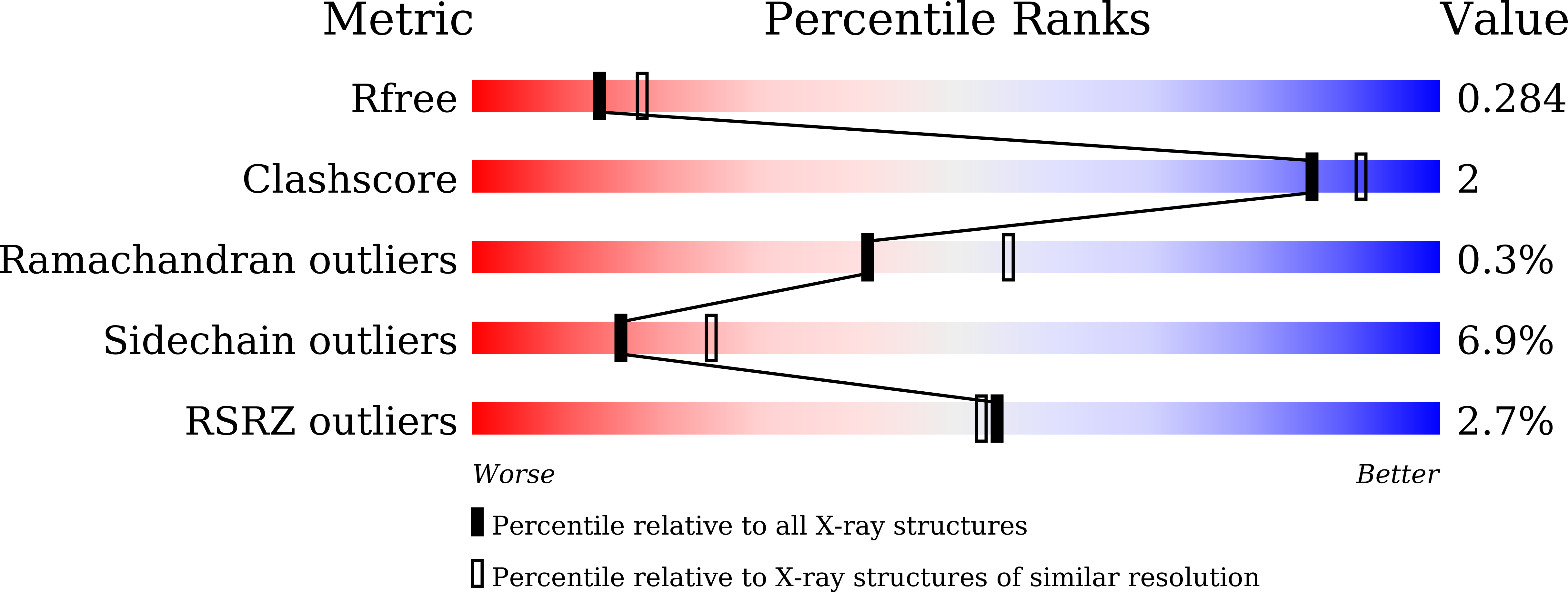
Experimental Data Snapshot
Entity ID: 1 | |||||
|---|---|---|---|---|---|
| Molecule | Chains | Sequence Length | Organism | Details | Image |
| Peptide deformylase | 168 | Pseudomonas aeruginosa | Mutation(s): 0 EC: 3.5.1.88 | 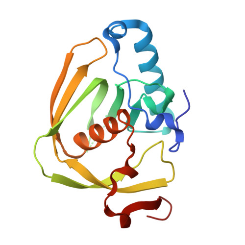 | |
UniProt | |||||
Find proteins for A0A071LDC0 (Pseudomonas aeruginosa) Explore A0A071LDC0 Go to UniProtKB: A0A071LDC0 | |||||
Entity Groups | |||||
| Sequence Clusters | 30% Identity50% Identity70% Identity90% Identity95% Identity100% Identity | ||||
| UniProt Group | A0A071LDC0 | ||||
Sequence AnnotationsExpand | |||||
| |||||
| Ligands 2 Unique | |||||
|---|---|---|---|---|---|
| ID | Chains | Name / Formula / InChI Key | 2D Diagram | 3D Interactions | |
| K1U (Subject of Investigation/LOI) Query on K1U | I [auth C] | (3R)-3-benzyl-4-oxo-4-[(2-oxo-2-phenylethyl)sulfanyl]butanoic acid C19 H18 O4 S XBDWLLDIBQGWMW-MRXNPFEDSA-N |  | ||
| NI Query on NI | E [auth A] F [auth B] G [auth B] H [auth C] J [auth D] | NICKEL (II) ION Ni VEQPNABPJHWNSG-UHFFFAOYSA-N |  | ||
| Length ( Å ) | Angle ( ˚ ) |
|---|---|
| a = 44.33 | α = 90 |
| b = 146.356 | β = 109.9 |
| c = 65.306 | γ = 90 |
| Software Name | Purpose |
|---|---|
| HKL-2000 | data scaling |
| REFMAC | refinement |
| HKL-2000 | data collection |
| HKL-2000 | data reduction |
| MOLREP | phasing |
RCSB PDB (citation) is hosted by
RCSB PDB is a member of the

