Structural Analysis of the Glycine Oxidase Homologue CmiS2 Reveals a Unique Substrate Recognition Mechanism for Formation of a beta-Amino Acid Starter Unit in Cremimycin Biosynthesis.
Kawasaki, D., Chisuga, T., Miyanaga, A., Kudo, F., Eguchi, T.(2019) Biochemistry 58: 2706-2709
- PubMed: 31154757
- DOI: https://doi.org/10.1021/acs.biochem.9b00444
- Primary Citation of Related Structures:
6J38, 6J39 - PubMed Abstract:
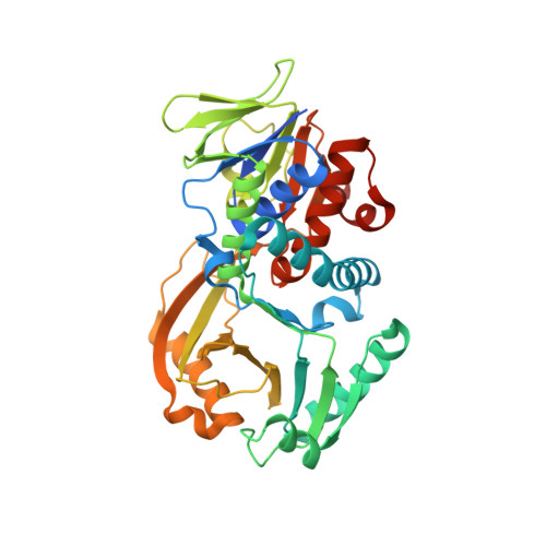
The flavin adenine dinucleotide-dependent oxidase CmiS2 catalyzes the oxidation of N-carboxymethyl-3-aminononanoic acid to produce a 3-aminononanoic acid starter unit for the biosynthesis of cremimycin, a macrolactam polyketide. Although the sequence of CmiS2 is similar with that of the well-characterized glycine oxidase ThiO, the chemical structure of the substrate of CmiS2 is different from that of ThiO substrate glycine. Here, we present the biochemical and structural characterization of CmiS2. Kinetic analysis revealed that CmiS2 has a strong preference for N-carboxymethyl-3-aminononanoic acid over other substrates such as N-carboxymethyl-3-aminobutanoic acid and glycine, suggesting that CmiS2 recognizes the nonanoic acid moiety of the substrate as well as the glycine moiety. We determined the crystal structure of CmiS2 in complex with a substrate analogue, namely, S-carboxymethyl-3-thiononanoic acid, which enabled the identification of key amino acid residues involved in substrate recognition. We discovered that Asn49, Arg243, and Arg334 interact with the carboxyl group of the nonanoic acid moiety, while Pro46, Leu52, and Ile335 recognize the alkyl chain of the nonanoic acid moiety via hydrophobic interaction. These residues are highly conserved in CmiS2 homologues involved in the biosynthesis of related macrolactam polyketides but are not conserved in glycine oxidases such as ThiO. These results suggest that CmiS2-type enzymes employ a distinct mechanism of substrate recognition for the synthesis of β-amino acids.
Organizational Affiliation:
Department of Chemistry , Tokyo Institute of Technology , 2-12-1 O-okayama , Meguro-ku, Tokyo 152-8551 , Japan.