6IP2

NSF-D1D2 part in the whole 20S complex


Experimental Data Snapshot

  • Method: ELECTRON MICROSCOPY
  • Resolution: 3.70 Å
  • Aggregation State: PARTICLE 
  • Reconstruction Method: SINGLE PARTICLE 

wwPDB Validation   3D Report Full Report


This is version 1.3 of the entry. See complete history


Literature

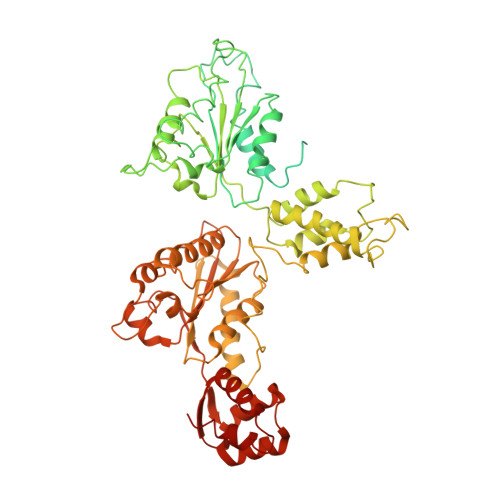
Mechanistic insights into the SNARE complex disassembly.

Huang, X.Sun, S.Wang, X.Fan, F.Zhou, Q.Lu, S.Cao, Y.Wang, Q.W.Dong, M.Q.Yao, J.Sui, S.F.

(2019) Sci Adv 5: eaau8164-eaau8164

  • DOI: https://doi.org/10.1126/sciadv.aau8164
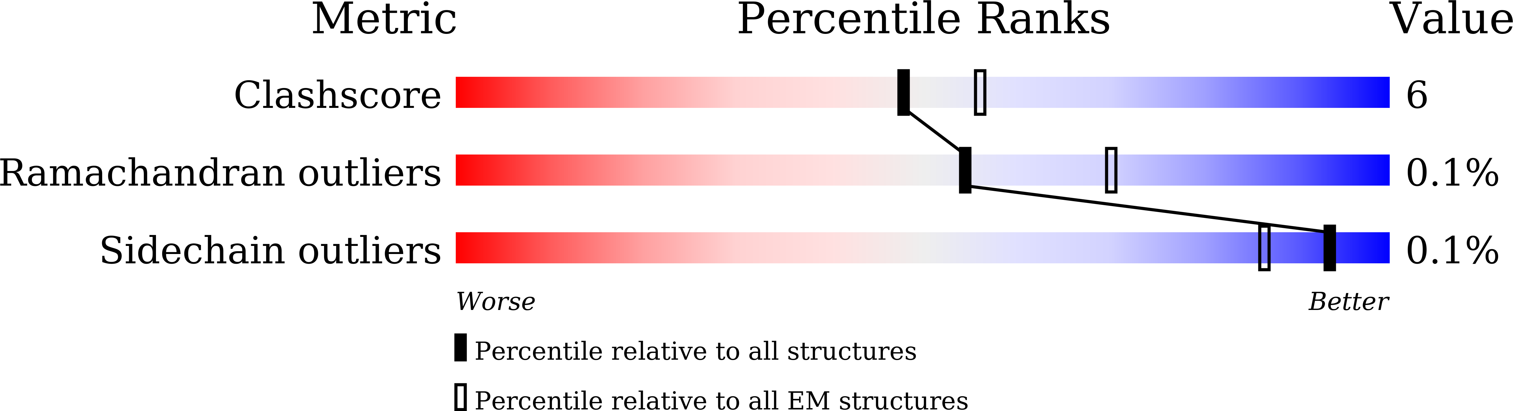
  • Primary Citation of Related Structures:  
    6IP1, 6IP2

  • PubMed Abstract: 

    NSF ( N -ethylmaleimide-sensitive factor) and α-SNAP (α-soluble NSF attachment protein) bind to the SNARE (soluble NSF attachment protein receptor) complex, the minimum machinery to mediate membrane fusion, to form a 20S complex, which disassembles the SNARE complex for reuse. We report the cryo-EM structures of the α-SNAP-SNARE subcomplex and the NSF-D1D2 domain in the 20S complex at 3.9- and 3.7-Å resolutions, respectively. Combined with the biochemical and electrophysiological analyses, we find that α-SNAPs use R116 through electrostatic interactions and L197 through hydrophobic interactions to apply force mainly on two positions of the VAMP protein to execute disassembly process. Furthermore, we define the interaction between the amino terminus of the SNARE helical bundle and the pore loop of the NSF-D1 domain and demonstrate its essential role as a potential anchor for SNARE complex disassembly. Our studies provide a rotation model of α-SNAP-mediated disassembly of the SNARE complex.


  • Organizational Affiliation

    State Key Laboratory of Membrane Biology, Beijing Advanced Innovation Center for Structural Biology, School of Life Sciences, Tsinghua University, Beijing 100084, China.


Macromolecules
Find similar proteins by:  (by identity cutoff)  |  3D Structure
Entity ID: 1
MoleculeChains Sequence LengthOrganismDetailsImage
Vesicle-fusing ATPase769Cricetulus griseusMutation(s): 0 
Gene Names: NSF
EC: 3.6.4.6
UniProt
Find proteins for P18708 (Cricetulus griseus)
Explore P18708 
Go to UniProtKB:  P18708
Entity Groups  
Sequence Clusters30% Identity50% Identity70% Identity90% Identity95% Identity100% Identity
UniProt GroupP18708
Sequence Annotations
Expand
  • Reference Sequence
Small Molecules
Ligands 1 Unique
IDChains Name / Formula / InChI Key2D Diagram3D Interactions
ATP
Query on ATP

Download Ideal Coordinates CCD File 
G [auth B]
H [auth B]
I [auth C]
J [auth C]
K [auth D]
G [auth B],
H [auth B],
I [auth C],
J [auth C],
K [auth D],
L [auth D],
M [auth E],
N [auth E],
O [auth F],
P [auth F],
Q [auth A],
R [auth A]
ADENOSINE-5'-TRIPHOSPHATE
C10 H16 N5 O13 P3
ZKHQWZAMYRWXGA-KQYNXXCUSA-N
Experimental Data & Validation

Experimental Data

  • Method: ELECTRON MICROSCOPY
  • Resolution: 3.70 Å
  • Aggregation State: PARTICLE 
  • Reconstruction Method: SINGLE PARTICLE 

Structure Validation

View Full Validation Report


Entry History 

Deposition Data

Revision History  (Full details and data files)

  • Version 1.0: 2019-04-24
    Type: Initial release
  • Version 1.1: 2019-05-01
    Changes: Data collection, Database references
  • Version 1.2: 2019-11-06
    Changes: Data collection, Other
  • Version 1.3: 2024-03-27
    Changes: Data collection, Database references