6ILU

Endolysin LysPBC5 CBD


Experimental Data Snapshot

  • Method: X-RAY DIFFRACTION
  • Resolution: 1.60 Å
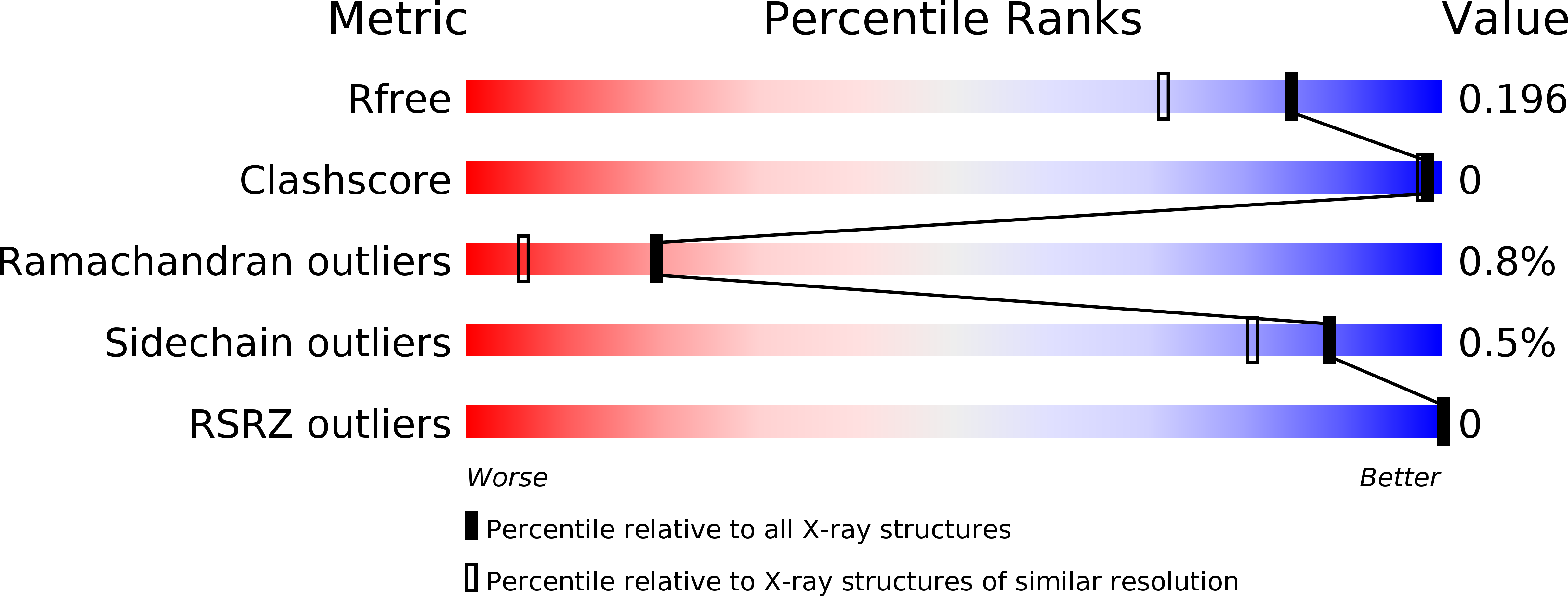
  • R-Value Free: 0.196 
  • R-Value Work: 0.167 
  • R-Value Observed: 0.168 

wwPDB Validation   3D Report Full Report


This is version 1.3 of the entry. See complete history


Literature

Structural Basis for Cell-Wall Recognition by Bacteriophage PBC5 Endolysin.

Lee, K.O.Kong, M.Kim, I.Bai, J.Cha, S.Kim, B.Ryu, K.S.Ryu, S.Suh, J.Y.

(2019) Structure 27: 1355-1365.e4

  • DOI: https://doi.org/10.1016/j.str.2019.07.001
  • Primary Citation of Related Structures:  
    6ILU

  • PubMed Abstract: 

    Phage endolysins are hydrolytic enzymes that cleave the bacterial cell wall during the lytic cycle. We isolated the bacteriophage PBC5 against Bacillus cereus, a major foodborne pathogen, and describe the molecular interaction between endolysin LysPBC5 and the host peptidoglycan structure. LysPBC5 has an N-terminal glycoside hydrolase 25 domain, and a C-terminal cell-wall binding domain (CBD) that is critical for specific cell-wall recognition and lysis. The crystal and solution structures of CBDs reveal tandem SH3b domains that are tightly engaged with each other. The CBD binds to the peptidoglycan in a bidentate manner via distal β sheet motifs with pseudo 2-fold symmetry, which can explain its high affinity and host specificity. The CBD primarily interacts with the glycan strand of the peptidoglycan layer instead of the peptide crosslink, implicating the tertiary structure of peptidoglycan as the recognition motif of endolysins.


  • Organizational Affiliation

    Department of Agricultural Biotechnology and Research Institute of Agriculture and Life Sciences, Seoul National University, Seoul 08826, South Korea; Protein Structure Research Team, Korea Basic Science Institute, 162 Yeongudanji-Ro, Ochang-Eup, Cheongju-Si, Chungcheongbuk-Do 28119, South Korea.


Macromolecules
Find similar proteins by:  (by identity cutoff)  |  3D Structure
Entity ID: 1
MoleculeChains Sequence LengthOrganismDetailsImage
Lysin
A, B
140Bacillus phage PBC5Mutation(s): 0 
Gene Names: PBC5_032
UniProt
Find proteins for A0A218KCJ1 (Bacillus phage PBC5)
Explore A0A218KCJ1 
Go to UniProtKB:  A0A218KCJ1
Entity Groups  
Sequence Clusters30% Identity50% Identity70% Identity90% Identity95% Identity100% Identity
UniProt GroupA0A218KCJ1
Sequence Annotations
Expand
  • Reference Sequence
Small Molecules
Ligands 2 Unique
IDChains Name / Formula / InChI Key2D Diagram3D Interactions
SO4
Query on SO4

Download Ideal Coordinates CCD File 
C [auth A]
D [auth A]
E [auth A]
F [auth A]
G [auth A]
C [auth A],
D [auth A],
E [auth A],
F [auth A],
G [auth A],
M [auth B],
N [auth B],
O [auth B]
SULFATE ION
O4 S
QAOWNCQODCNURD-UHFFFAOYSA-L
EDO
Query on EDO

Download Ideal Coordinates CCD File 
H [auth A]
I [auth A]
J [auth A]
K [auth A]
L [auth A]
H [auth A],
I [auth A],
J [auth A],
K [auth A],
L [auth A],
P [auth B],
Q [auth B],
R [auth B],
S [auth B],
T [auth B],
U [auth B],
V [auth B]
1,2-ETHANEDIOL
C2 H6 O2
LYCAIKOWRPUZTN-UHFFFAOYSA-N
Experimental Data & Validation

Experimental Data

  • Method: X-RAY DIFFRACTION
  • Resolution: 1.60 Å
  • R-Value Free: 0.196 
  • R-Value Work: 0.167 
  • R-Value Observed: 0.168 
  • Space Group: I 2 2 2
Unit Cell:
Length ( Å )Angle ( ˚ )
a = 81.823α = 90
b = 107.514β = 90
c = 117.524γ = 90
Software Package:
Software NamePurpose
PHENIXrefinement
HKL-2000data reduction
HKL-2000data scaling
SOLVEphasing

Structure Validation

View Full Validation Report


Entry History 

Deposition Data

Revision History  (Full details and data files)

  • Version 1.0: 2019-07-31
    Type: Initial release
  • Version 1.1: 2019-08-07
    Changes: Data collection, Derived calculations
  • Version 1.2: 2020-02-12
    Changes: Database references
  • Version 1.3: 2024-03-27
    Changes: Data collection, Database references