6I7E

Plasmodium falciparum Myosin A, Pre-powerstroke


Experimental Data Snapshot

  • Method: X-RAY DIFFRACTION
  • Resolution: 3.49 Å
  • R-Value Free: 0.233 
  • R-Value Work: 0.193 
  • R-Value Observed: 0.195 

wwPDB Validation   3D Report Full Report


Ligand Structure Quality Assessment 


This is version 1.1 of the entry. See complete history


Literature

Plasmodium myosin A drives parasite invasion by an atypical force generating mechanism.

Robert-Paganin, J.Robblee, J.P.Auguin, D.Blake, T.C.A.Bookwalter, C.S.Krementsova, E.B.Moussaoui, D.Previs, M.J.Jousset, G.Baum, J.Trybus, K.M.Houdusse, A.

(2019) Nat Commun 10: 3286-3286

  • DOI: https://doi.org/10.1038/s41467-019-11120-0
  • Primary Citation of Related Structures:  
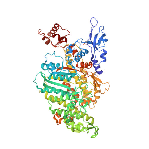
    6I7D, 6I7E

  • PubMed Abstract: 

    Plasmodium parasites are obligate intracellular protozoa and causative agents of malaria, responsible for half a million deaths each year. The lifecycle progression of the parasite is reliant on cell motility, a process driven by myosin A, an unconventional single-headed class XIV molecular motor. Here we demonstrate that myosin A from Plasmodium falciparum (PfMyoA) is critical for red blood cell invasion. Further, using a combination of X-ray crystallography, kinetics, and in vitro motility assays, we elucidate the non-canonical interactions that drive this motor's function. We show that PfMyoA motor properties are tuned by heavy chain phosphorylation (Ser19), with unphosphorylated PfMyoA exhibiting enhanced ensemble force generation at the expense of speed. Regulated phosphorylation may therefore optimize PfMyoA for enhanced force generation during parasite invasion or for fast motility during dissemination. The three PfMyoA crystallographic structures presented here provide a blueprint for discovery of specific inhibitors designed to prevent parasite infection.


  • Organizational Affiliation

    Structural Motility, UMR 144 CNRS/Curie Institute, 26 rue d'ulm, 75258, Paris cedex 05, France.


Macromolecules
Find similar proteins by:  (by identity cutoff)  |  3D Structure
Entity ID: 1
MoleculeChains Sequence LengthOrganismDetailsImage
Myosin-A768Plasmodium falciparum 3D7Mutation(s): 0 
Gene Names: PF13_0233
UniProt
Find proteins for Q8IDR3 (Plasmodium falciparum (isolate 3D7))
Explore Q8IDR3 
Go to UniProtKB:  Q8IDR3
Entity Groups  
Sequence Clusters30% Identity50% Identity70% Identity90% Identity95% Identity100% Identity
UniProt GroupQ8IDR3
Sequence Annotations
Expand
  • Reference Sequence
Small Molecules
Modified Residues  1 Unique
IDChains TypeFormula2D DiagramParent
SEP
Query on SEP
A
L-PEPTIDE LINKINGC3 H8 N O6 PSER
Experimental Data & Validation

Experimental Data

  • Method: X-RAY DIFFRACTION
  • Resolution: 3.49 Å
  • R-Value Free: 0.233 
  • R-Value Work: 0.193 
  • R-Value Observed: 0.195 
  • Space Group: P 61 2 2
Unit Cell:
Length ( Å )Angle ( ˚ )
a = 197.46α = 90
b = 197.46β = 90
c = 178.144γ = 120
Software Package:
Software NamePurpose
BUSTERrefinement
XDSdata reduction
XDSdata scaling
PHASERphasing

Structure Validation

View Full Validation Report


Ligand Structure Quality Assessment 


Entry History & Funding Information

Deposition Data


Funding OrganizationLocationGrant Number
Human Frontier Science ProgramUnited StatesRGY0066/2016
Wellcome TrustUnited Kingdom109007/Z/15/A
Wellcome TrustUnited Kingdom100993/Z/13/Z

Revision History  (Full details and data files)

  • Version 1.0: 2019-08-07
    Type: Initial release
  • Version 1.1: 2024-01-24
    Changes: Data collection, Database references, Refinement description