6HLP

Crystal structure of the Neurokinin 1 receptor in complex with the small molecule antagonist Netupitant


Experimental Data Snapshot

  • Method: X-RAY DIFFRACTION
  • Resolution: 2.20 Å
  • R-Value Free: 0.225 
  • R-Value Work: 0.203 
  • R-Value Observed: 0.204 

wwPDB Validation   3D Report Full Report


Ligand Structure Quality Assessment 


This is version 1.1 of the entry. See complete history


Literature

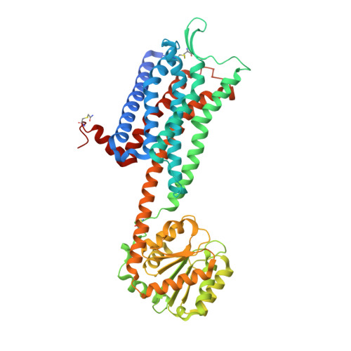
Crystal structures of the human neurokinin 1 receptor in complex with clinically used antagonists.

Schoppe, J.Ehrenmann, J.Klenk, C.Rucktooa, P.Schutz, M.Dore, A.S.Pluckthun, A.

(2019) Nat Commun 10: 17-17

  • DOI: https://doi.org/10.1038/s41467-018-07939-8
  • Primary Citation of Related Structures:  
    6HLL, 6HLO, 6HLP

  • PubMed Abstract: 

    Neurokinins (or tachykinins) are peptides that modulate a wide variety of human physiology through the neurokinin G protein-coupled receptor family, implicated in a diverse array of pathological processes. Here we report high-resolution crystal structures of the human NK 1 receptor (NK 1 R) bound to two small-molecule antagonist therapeutics - aprepitant and netupitant and the progenitor antagonist CP-99,994. The structures reveal the detailed interactions between clinically approved antagonists and NK 1 R, which induce a distinct receptor conformation resulting in an interhelical hydrogen-bond network that cross-links the extracellular ends of helices V and VI. Furthermore, the high-resolution details of NK 1 R bound to netupitant establish a structural rationale for the lack of basal activity in NK 1 R. Taken together, these co-structures provide a comprehensive structural basis of NK 1 R antagonism and will facilitate the design of new therapeutics targeting the neurokinin receptor family.


  • Organizational Affiliation

    Department of Biochemistry, University of Zürich, Winterthurerstrasse 190, CH-8057, Zürich, Switzerland.


Macromolecules
Find similar proteins by:  (by identity cutoff)  |  3D Structure
Entity ID: 1
MoleculeChains Sequence LengthOrganismDetailsImage
Substance-P receptor,Substance-P receptor520Homo sapiensPyrococcus abyssi GE5Mutation(s): 7 
Gene Names: TACR1NK1RTAC1R
Membrane Entity: Yes 
UniProt & NIH Common Fund Data Resources
Find proteins for Q9V2J8 (Pyrococcus abyssi (strain GE5 / Orsay))
Explore Q9V2J8 
Go to UniProtKB:  Q9V2J8
Find proteins for P25103 (Homo sapiens)
Explore P25103 
Go to UniProtKB:  P25103
PHAROS:  P25103
GTEx:  ENSG00000115353 
Entity Groups  
Sequence Clusters30% Identity50% Identity70% Identity90% Identity95% Identity100% Identity
UniProt GroupsQ9V2J8P25103
Sequence Annotations
Expand
  • Reference Sequence
Small Molecules
Ligands 5 Unique
IDChains Name / Formula / InChI Key2D Diagram3D Interactions
GAW
Query on GAW

Download Ideal Coordinates CCD File 
B [auth A]2-[3,5-bis(trifluoromethyl)phenyl]-~{N},2-dimethyl-~{N}-[4-(2-methylphenyl)-6-(4-methylpiperazin-1-yl)pyridin-3-yl]propanamide
C30 H32 F6 N4 O
WAXQNWCZJDTGBU-UHFFFAOYSA-N
OLC
Query on OLC

Download Ideal Coordinates CCD File 
M [auth A]
N [auth A]
NA [auth A]
O [auth A]
OA [auth A]
M [auth A],
N [auth A],
NA [auth A],
O [auth A],
OA [auth A],
P [auth A],
Q [auth A]
(2R)-2,3-dihydroxypropyl (9Z)-octadec-9-enoate
C21 H40 O4
RZRNAYUHWVFMIP-GDCKJWNLSA-N
OLA
Query on OLA

Download Ideal Coordinates CCD File 
D [auth A]
E [auth A]
F [auth A]
G [auth A]
H [auth A]
D [auth A],
E [auth A],
F [auth A],
G [auth A],
H [auth A],
I [auth A],
J [auth A],
K [auth A],
L [auth A]
OLEIC ACID
C18 H34 O2
ZQPPMHVWECSIRJ-KTKRTIGZSA-N
CIT
Query on CIT

Download Ideal Coordinates CCD File 
C [auth A]CITRIC ACID
C6 H8 O7
KRKNYBCHXYNGOX-UHFFFAOYSA-N
PEG
Query on PEG

Download Ideal Coordinates CCD File 
AA [auth A]
BA [auth A]
CA [auth A]
DA [auth A]
EA [auth A]
AA [auth A],
BA [auth A],
CA [auth A],
DA [auth A],
EA [auth A],
FA [auth A],
GA [auth A],
HA [auth A],
IA [auth A],
JA [auth A],
KA [auth A],
LA [auth A],
MA [auth A],
PA [auth A],
QA [auth A],
R [auth A],
S [auth A],
T [auth A],
U [auth A],
V [auth A],
W [auth A],
X [auth A],
Y [auth A],
Z [auth A]
DI(HYDROXYETHYL)ETHER
C4 H10 O3
MTHSVFCYNBDYFN-UHFFFAOYSA-N
Modified Residues  1 Unique
IDChains TypeFormula2D DiagramParent
YCM
Query on YCM
A
L-PEPTIDE LINKINGC5 H10 N2 O3 SCYS
Binding Affinity Annotations 
IDSourceBinding Affinity
GAW BindingDB:  6HLP Ki: 0.95 (nM) from 1 assay(s)
IC50: 0.95 (nM) from 1 assay(s)
Experimental Data & Validation

Experimental Data

  • Method: X-RAY DIFFRACTION
  • Resolution: 2.20 Å
  • R-Value Free: 0.225 
  • R-Value Work: 0.203 
  • R-Value Observed: 0.204 
  • Space Group: P 21 21 21
Unit Cell:
Length ( Å )Angle ( ˚ )
a = 61.658α = 90
b = 76.571β = 90
c = 166.035γ = 90
Software Package:
Software NamePurpose
PHENIXrefinement
XDSdata reduction
Aimlessdata scaling
PHASERphasing

Structure Validation

View Full Validation Report


Ligand Structure Quality Assessment 


Entry History & Funding Information

Deposition Data


Funding OrganizationLocationGrant Number
Swiss National Science FoundationSwitzerland31003A_153143

Revision History  (Full details and data files)

  • Version 1.0: 2019-01-16
    Type: Initial release
  • Version 1.1: 2024-01-24
    Changes: Data collection, Database references, Derived calculations, Refinement description