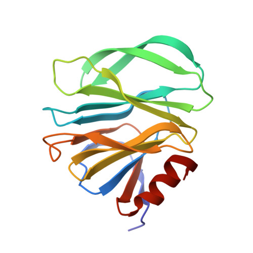
6H9Y

Unraveling the role of the secretor antigen in human rotavirus attachment to histo-blood group antigens


Experimental Data Snapshot

  • Method: X-RAY DIFFRACTION
  • Resolution: 1.31 Å
  • R-Value Free: 0.209 
  • R-Value Work: 0.181 
  • R-Value Observed: 0.182 

wwPDB Validation   3D Report Full Report


This is version 2.1 of the entry. See complete history


Literature

Unraveling the role of the secretor antigen in human rotavirus attachment to histo-blood group antigens.

Gozalbo-Rovira, R.Ciges-Tomas, J.R.Vila-Vicent, S.Buesa, J.Santiso-Bellon, C.Monedero, V.Yebra, M.J.Marina, A.Rodriguez-Diaz, J.

(2019) PLoS Pathog 15: e1007865-e1007865

  • DOI: https://doi.org/10.1371/journal.ppat.1007865
  • Primary Citation of Related Structures:  
    6H9W, 6H9Y, 6H9Z, 6HA0

  • PubMed Abstract: 

    Rotavirus is the leading agent causing acute gastroenteritis in young children, with the P[8] genotype accounting for more than 80% of infections in humans. The molecular bases for binding of the VP8* domain from P[8] VP4 spike protein to its cellular receptor, the secretory H type-1 antigen (Fuc-α1,2-Gal-β1,3-GlcNAc; H1), and to its precursor lacto-N-biose (Gal-β1,3-GlcNAc; LNB) have been determined. The resolution of P[8] VP8* crystal structures in complex with H1 antigen and LNB and site-directed mutagenesis experiments revealed that both glycans bind to the P[8] VP8* protein through a binding pocket shared with other members of the P[II] genogroup (i.e.: P[4], P[6] and P[19]). Our results show that the L-fucose moiety from H1 only displays indirect contacts with P[8] VP8*. However, the induced conformational changes in the LNB moiety increase the ligand affinity by two-fold, as measured by surface plasmon resonance (SPR), providing a molecular explanation for the different susceptibility to rotavirus infection between secretor and non-secretor individuals. The unexpected interaction of P[8] VP8* with LNB, a building block of type-1 human milk oligosaccharides, resulted in inhibition of rotavirus infection, highlighting the role and possible application of this disaccharide as an antiviral. While key amino acids in the H1/LNB binding pocket were highly conserved in members of the P[II] genogroup, differences were found in ligand affinities among distinct P[8] genetic lineages. The variation in affinities were explained by subtle structural differences induced by amino acid changes in the vicinity of the binding pocket, providing a fine-tuning mechanism for glycan binding in P[8] rotavirus.


  • Organizational Affiliation

    Departament of Microbiology, Faculty of Medicine, University of Valencia, Valencia, Spain.


Macromolecules
Find similar proteins by:  (by identity cutoff)  |  3D Structure
Entity ID: 1
MoleculeChains Sequence LengthOrganismDetailsImage
Outer capsid protein VP4
A, B
162Human rotavirus AMutation(s): 0 
Gene Names: VP4
UniProt
Find proteins for Q0PRF2 (Human rotavirus A)
Explore Q0PRF2 
Go to UniProtKB:  Q0PRF2
Entity Groups  
Sequence Clusters30% Identity50% Identity70% Identity90% Identity95% Identity100% Identity
UniProt GroupQ0PRF2
Sequence Annotations
Expand
  • Reference Sequence
Oligosaccharides

Help

Entity ID: 2
MoleculeChains Length2D Diagram Glycosylation3D Interactions
beta-D-galactopyranose-(1-3)-2-acetamido-2-deoxy-beta-D-glucopyranose
C
2N/A
Glycosylation Resources
GlyTouCan:  G00056MO
GlyCosmos:  G00056MO
GlyGen:  G00056MO
Small Molecules
Ligands 1 Unique
IDChains Name / Formula / InChI Key2D Diagram3D Interactions
BTB
Query on BTB

Download Ideal Coordinates CCD File 
D [auth B]2-[BIS-(2-HYDROXY-ETHYL)-AMINO]-2-HYDROXYMETHYL-PROPANE-1,3-DIOL
C8 H19 N O5
OWMVSZAMULFTJU-UHFFFAOYSA-N
Experimental Data & Validation

Experimental Data

  • Method: X-RAY DIFFRACTION
  • Resolution: 1.31 Å
  • R-Value Free: 0.209 
  • R-Value Work: 0.181 
  • R-Value Observed: 0.182 
  • Space Group: P 1 21 1
Unit Cell:
Length ( Å )Angle ( ˚ )
a = 38.988α = 90
b = 54.725β = 97.92
c = 67.433γ = 90
Software Package:
Software NamePurpose
XDSdata reduction
Aimlessdata scaling
REFMACrefinement
PDB_EXTRACTdata extraction
PHASERphasing

Structure Validation

View Full Validation Report


Entry History & Funding Information

Deposition Data


Funding OrganizationLocationGrant Number
Spanish Ministry of Economy and CompetitivenessSpainBIO2016-78571-P

Revision History  (Full details and data files)

  • Version 1.0: 2019-06-12
    Type: Initial release
  • Version 1.1: 2019-07-03
    Changes: Data collection, Database references
  • Version 2.0: 2020-07-29
    Type: Remediation
    Reason: Carbohydrate remediation
    Changes: Atomic model, Data collection, Derived calculations, Structure summary
  • Version 2.1: 2024-01-17
    Changes: Data collection, Database references, Derived calculations, Refinement description, Structure summary