6H76

SiaP in complex with Neu5Ac (RT)


Experimental Data Snapshot

  • Method: X-RAY DIFFRACTION
  • Resolution: 1.50 Å
  • R-Value Free: 0.155 
  • R-Value Work: 0.112 
  • R-Value Observed: 0.114 

wwPDB Validation   3D Report Full Report


Ligand Structure Quality Assessment 


This is version 1.3 of the entry. See complete history


Literature

Water Networks Can Determine the Affinity of Ligand Binding to Proteins.

Darby, J.F.Hopkins, A.P.Shimizu, S.Roberts, S.M.Brannigan, J.A.Turkenburg, J.P.Thomas, G.H.Hubbard, R.E.Fischer, M.

(2019) J Am Chem Soc 141: 15818-15826

  • DOI: https://doi.org/10.1021/jacs.9b06275
  • Primary Citation of Related Structures:  
    2V4C, 2WX9, 2WYK, 2WYP, 2XA5, 6H75, 6H76

  • PubMed Abstract: 

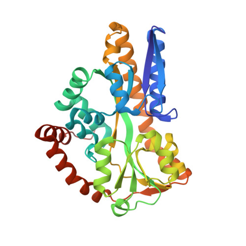
    Solvent organization is a key but underexploited contributor to the thermodynamics of protein-ligand recognition, with implications for ligand discovery, drug resistance, and protein engineering. Here, we explore the contribution of solvent to ligand binding in the Haemophilus influenzae virulence protein SiaP. By introducing a single mutation without direct ligand contacts, we observed a >1000-fold change in sialic acid binding affinity. Crystallographic and calorimetric data of wild-type and mutant SiaP showed that this change results from an enthalpically unfavorable perturbation of the solvent network. This disruption is reflected by changes in the normalized atomic displacement parameters of crystallographic water molecules. In SiaP's enclosed cavity, relative differences in water-network dynamics serve as a simple predictor of changes in the free energy of binding upon changing protein, ligand, or both. This suggests that solvent structure is an evolutionary constraint on protein sequence that contributes to ligand affinity and selectivity.


  • Organizational Affiliation

    Demuris Ltd., The Biosphere , Draymans Way, Newcastle Helix , Newcastle upon Tyne NE4 5BX , United Kingdom.


Macromolecules
Find similar proteins by:  (by identity cutoff)  |  3D Structure
Entity ID: 1
MoleculeChains Sequence LengthOrganismDetailsImage
Sialic acid-binding periplasmic protein SiaP312Haemophilus influenzae Rd KW20Mutation(s): 0 
Gene Names: siaPHI_0146
UniProt
Find proteins for P44542 (Haemophilus influenzae (strain ATCC 51907 / DSM 11121 / KW20 / Rd))
Explore P44542 
Go to UniProtKB:  P44542
Entity Groups  
Sequence Clusters30% Identity50% Identity70% Identity90% Identity95% Identity100% Identity
UniProt GroupP44542
Sequence Annotations
Expand
  • Reference Sequence
Experimental Data & Validation

Experimental Data

  • Method: X-RAY DIFFRACTION
  • Resolution: 1.50 Å
  • R-Value Free: 0.155 
  • R-Value Work: 0.112 
  • R-Value Observed: 0.114 
  • Space Group: P 21 21 21
Unit Cell:
Length ( Å )Angle ( ˚ )
a = 47.788α = 90
b = 75.31β = 90
c = 87.774γ = 90
Software Package:
Software NamePurpose
REFMACrefinement
DENZOdata reduction
SCALEPACKdata scaling
ACORNphasing

Structure Validation

View Full Validation Report


Ligand Structure Quality Assessment 


Entry History & Funding Information

Deposition Data


Funding OrganizationLocationGrant Number
Biotechnology and Biological Sciences Research CouncilUnited KingdomBBC509807

Revision History  (Full details and data files)

  • Version 1.0: 2019-08-14
    Type: Initial release
  • Version 1.1: 2019-09-25
    Changes: Data collection, Database references
  • Version 1.2: 2019-10-16
    Changes: Data collection, Database references
  • Version 1.3: 2020-07-29
    Type: Remediation
    Reason: Carbohydrate remediation
    Changes: Data collection, Derived calculations, Structure summary