6GDT

Crystal structure of exo-glucosidase/glucosaminidase VC0615 from Vibrio Cholerae


Experimental Data Snapshot

  • Method: X-RAY DIFFRACTION
  • Resolution: 3.17 Å
  • R-Value Free: 0.280 
  • R-Value Work: 0.235 
  • R-Value Observed: 0.237 

wwPDB Validation   3D Report Full Report


This is version 1.1 of the entry. See complete history


Literature

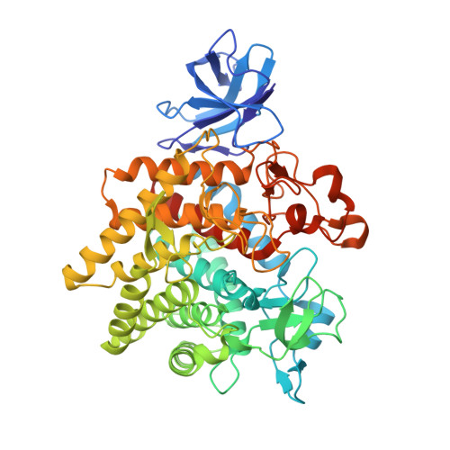
Structure of the GH9 glucosidase/glucosaminidase from Vibrio cholerae.

Wu, L.Davies, G.J.

(2018) Acta Crystallogr F Struct Biol Commun 74: 512-523

  • DOI: https://doi.org/10.1107/S2053230X18011019
  • Primary Citation of Related Structures:  
    6GDT

  • PubMed Abstract: 

    Glycoside hydrolase family 9 (GH9) of carbohydrate-processing enzymes primarily consists of inverting endoglucanases. A subgroup of GH9 enzymes are believed to act as exo-glucosidases or exo-glucosaminidases, with many being found in organisms of the family Vibrionaceae, where they are proposed to function within the chitin-catabolism pathway. Here, it is shown that the GH9 enzyme from the pathogen Vibrio cholerae (hereafter referred to as VC0615) is active on both chitosan-derived and β-glucoside substrates. The structure of VC0615 at 3.17 Å resolution is reported from a crystal form with poor diffraction and lattice disorder. VC0615 was highly refractory to crystallization efforts, with crystals only appearing using a high protein concentration under conditions containing the precipitant poly-γ-glutamic acid (PGA). The structure is highly mobile within the crystal lattice, which is likely to reflect steric clashes between symmetry molecules which destabilize crystal packing. The overall tertiary structure of VC0615 is well resolved even at 3.17 Å resolution, which has allowed the structural basis for the exo-glucosidase/glucosaminidase activity of this enzyme to be investigated.


  • Organizational Affiliation

    York Structural Biology Laboratory, Department of Chemistry, The University of York, York YO10 5DD, England.


Macromolecules
Find similar proteins by:  (by identity cutoff)  |  3D Structure
Entity ID: 1
MoleculeChains Sequence LengthOrganismDetailsImage
Endoglucanase-related protein
A, B, C, D, E
582Vibrio cholerae O1 biovar El Tor str. N16961Mutation(s): 0 
Gene Names: VC_0615
UniProt
Find proteins for Q9KUA8 (Vibrio cholerae serotype O1 (strain ATCC 39315 / El Tor Inaba N16961))
Explore Q9KUA8 
Go to UniProtKB:  Q9KUA8
Entity Groups  
Sequence Clusters30% Identity50% Identity70% Identity90% Identity95% Identity100% Identity
UniProt GroupQ9KUA8
Sequence Annotations
Expand
  • Reference Sequence
Small Molecules
Ligands 1 Unique
IDChains Name / Formula / InChI Key2D Diagram3D Interactions
EDO
Query on EDO

Download Ideal Coordinates CCD File 
F [auth C]1,2-ETHANEDIOL
C2 H6 O2
LYCAIKOWRPUZTN-UHFFFAOYSA-N
Experimental Data & Validation

Experimental Data

  • Method: X-RAY DIFFRACTION
  • Resolution: 3.17 Å
  • R-Value Free: 0.280 
  • R-Value Work: 0.235 
  • R-Value Observed: 0.237 
  • Space Group: P 32 2 1
Unit Cell:
Length ( Å )Angle ( ˚ )
a = 234.899α = 90
b = 234.899β = 90
c = 129.421γ = 120
Software Package:
Software NamePurpose
REFMACrefinement
XDSdata reduction
Aimlessdata scaling
PHASERphasing

Structure Validation

View Full Validation Report


Entry History & Funding Information

Deposition Data


Funding OrganizationLocationGrant Number
European Research CouncilUnited KingdomERC-2012-AdG-32294

Revision History  (Full details and data files)

  • Version 1.0: 2018-08-15
    Type: Initial release
  • Version 1.1: 2024-01-17
    Changes: Data collection, Database references, Refinement description