6FLF

Deoxyguanylosuccinate synthase (DgsS) structure at 1.33 Angstrom resolution.


Experimental Data Snapshot

  • Method: X-RAY DIFFRACTION
  • Resolution: 1.33 Å
  • R-Value Free: 0.226 
  • R-Value Work: 0.200 
  • R-Value Observed: 0.201 

wwPDB Validation   3D Report Full Report


This is version 1.2 of the entry. See complete history


Literature

A third purine biosynthetic pathway encoded by aminoadenine-based viral DNA genomes.

Sleiman, D.Garcia, P.S.Lagune, M.Loc'h, J.Haouz, A.Taib, N.Rothlisberger, P.Gribaldo, S.Marliere, P.Kaminski, P.A.

(2021) Science 372: 516-520

  • DOI: https://doi.org/10.1126/science.abe6494
  • Primary Citation of Related Structures:  
    6FLF, 6FM0, 6FM1, 6TNH

  • PubMed Abstract: 

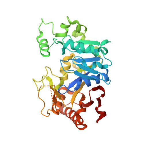
    Cells have two purine pathways that synthesize adenine and guanine ribonucleotides from phosphoribose via inosylate. A chemical hybrid between adenine and guanine, 2-aminoadenine (Z), replaces adenine in the DNA of the cyanobacterial virus S-2L. We show that S-2L and Vibrio phage PhiVC8 encode a third purine pathway catalyzed by PurZ, a distant paralog of succinoadenylate synthase (PurA), the enzyme condensing aspartate and inosylate in the adenine pathway. PurZ condenses aspartate with deoxyguanylate into dSMP (N6-succino-2-amino-2'-deoxyadenylate), which undergoes defumarylation and phosphorylation to give dZTP (2-amino-2'-deoxyadenosine-5'-triphosphate), a substrate for the phage DNA polymerase. Crystallography and phylogenetics analyses indicate a close relationship between phage PurZ and archaeal PurA enzymes. Our work elucidates the biocatalytic innovation that remodeled a DNA building block beyond canonical molecular biology.


  • Organizational Affiliation

    Biology of Gram-Positive Pathogens, Institut Pasteur, CNRS-UMR 2001, Paris, France.


Macromolecules
Find similar proteins by:  (by identity cutoff)  |  3D Structure
Entity ID: 1
MoleculeChains Sequence LengthOrganismDetailsImage
Adenylosuccinate synthetase363Vibrio phage phiVC8Mutation(s): 0 
Gene Names: phiVC8_p27
UniProt
Find proteins for G3FFN6 (Vibrio phage phiVC8)
Explore G3FFN6 
Go to UniProtKB:  G3FFN6
Entity Groups  
Sequence Clusters30% Identity50% Identity70% Identity90% Identity95% Identity100% Identity
UniProt GroupG3FFN6
Sequence Annotations
Expand
  • Reference Sequence
Experimental Data & Validation

Experimental Data

  • Method: X-RAY DIFFRACTION
  • Resolution: 1.33 Å
  • R-Value Free: 0.226 
  • R-Value Work: 0.200 
  • R-Value Observed: 0.201 
  • Space Group: C 1 2 1
Unit Cell:
Length ( Å )Angle ( ˚ )
a = 90.92α = 90
b = 59.48β = 102.67
c = 73.98γ = 90
Software Package:
Software NamePurpose
BUSTERrefinement
XDSdata reduction
SCALAdata scaling
PHASERphasing

Structure Validation

View Full Validation Report


Entry History 

Deposition Data

Revision History  (Full details and data files)

  • Version 1.0: 2019-06-12
    Type: Initial release
  • Version 1.1: 2019-06-26
    Changes: Data collection, Structure summary
  • Version 1.2: 2021-06-02
    Changes: Database references