6F5K

Crystal structure of laccase from Myceliophthora thermophila


Experimental Data Snapshot

  • Method: X-RAY DIFFRACTION
  • Resolution: 1.62 Å
  • R-Value Free: 0.189 
  • R-Value Work: 0.150 
  • R-Value Observed: 0.152 

wwPDB Validation   3D Report Full Report


Ligand Structure Quality Assessment 


This is version 3.1 of the entry. See complete history


Literature

A comparative structural analysis of the surface properties of asco-laccases.

Ernst, H.A.Jorgensen, L.J.Bukh, C.Piontek, K.Plattner, D.A.Ostergaard, L.H.Larsen, S.Bjerrum, M.J.

(2018) PLoS One 13: e0206589-e0206589

  • DOI: https://doi.org/10.1371/journal.pone.0206589
  • Primary Citation of Related Structures:  
    6F5K

  • PubMed Abstract: 

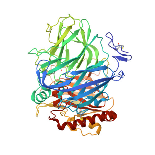
    Laccases of different biological origins have been widely investigated and these studies have elucidated fundamentals of the generic catalytic mechanism. However, other features such as surface properties and residues located away from the catalytic centres may also have impact on enzyme function. Here we present the crystal structure of laccase from Myceliophthora thermophila (MtL) to a resolution of 1.62 Å together with a thorough structural comparison with other members of the CAZy family AA1_3 that comprises fungal laccases from ascomycetes. The recombinant protein produced in A. oryzae has a molecular mass of 75 kDa, a pI of 4.2 and carries 13.5 kDa N-linked glycans. In the crystal, MtL forms a dimer with the phenolic substrate binding pocket blocked, suggesting that the active form of the enzyme is monomeric. Overall, the MtL structure conforms with the canonical fold of fungal laccases as well as the features specific for the asco-laccases. However, the structural comparisons also reveal significant variations within this taxonomic subgroup. Notable differences in the T1-Cu active site topology and polar motifs imply molecular evolution to serve different functional roles. Very few surface residues are conserved and it is noticeable that they encompass residues that interact with the N-glycans and/or are located at domain interfaces. The N-glycosylation sites are surprisingly conserved among asco-laccases and in most cases the glycan displays extensive interactions with the protein. In particular, the glycans at Asn88 and Asn210 appear to have evolved as an integral part of the asco-laccase structure. An uneven distribution of the carbohydrates around the enzyme give unique properties to a distinct part of the surface of the asco-laccases which may have implication for laccase function-in particular towards large substrates.


  • Organizational Affiliation

    Department of Chemistry, University of Copenhagen, Copenhagen, Denmark.


Macromolecules
Find similar proteins by:  (by identity cutoff)  |  3D Structure
Entity ID: 1
MoleculeChains Sequence LengthOrganismDetailsImage
Extracellular laccase, lcc1559Thermothelomyces thermophilusMutation(s): 0 
Gene Names: lcc1MYCTH_51627
UniProt
Find proteins for G2QG31 (Thermothelomyces thermophilus (strain ATCC 42464 / BCRC 31852 / DSM 1799))
Explore G2QG31 
Go to UniProtKB:  G2QG31
Entity Groups  
Sequence Clusters30% Identity50% Identity70% Identity90% Identity95% Identity100% Identity
UniProt GroupG2QG31
Sequence Annotations
Expand
  • Reference Sequence
Oligosaccharides

Help

Entity ID: 2
MoleculeChains Length2D Diagram Glycosylation3D Interactions
alpha-D-mannopyranose-(1-3)-alpha-D-mannopyranose-(1-6)-beta-D-mannopyranose-(1-4)-2-acetamido-2-deoxy-beta-D-glucopyranose-(1-4)-2-acetamido-2-deoxy-beta-D-glucopyranose
B
5N-Glycosylation
Glycosylation Resources
GlyTouCan:  G10756ZZ
GlyCosmos:  G10756ZZ
GlyGen:  G10756ZZ
Entity ID: 3
MoleculeChains Length2D Diagram Glycosylation3D Interactions
2-acetamido-2-deoxy-beta-D-glucopyranose-(1-4)-2-acetamido-2-deoxy-beta-D-glucopyranose
C
2N-Glycosylation
Glycosylation Resources
GlyTouCan:  G42666HT
GlyCosmos:  G42666HT
GlyGen:  G42666HT
Entity ID: 4
MoleculeChains Length2D Diagram Glycosylation3D Interactions
alpha-D-mannopyranose-(1-2)-alpha-D-mannopyranose-(1-2)-alpha-D-mannopyranose-(1-3)-beta-D-mannopyranose-(1-4)-2-acetamido-2-deoxy-beta-D-glucopyranose-(1-4)-2-acetamido-2-deoxy-beta-D-glucopyranose
D
6N-Glycosylation
Glycosylation Resources
GlyTouCan:  G01760ZU
GlyCosmos:  G01760ZU
GlyGen:  G01760ZU
Entity ID: 5
MoleculeChains Length2D Diagram Glycosylation3D Interactions
beta-D-mannopyranose-(1-4)-2-acetamido-2-deoxy-beta-D-glucopyranose-(1-4)-2-acetamido-2-deoxy-beta-D-glucopyranose
E, F
3N-Glycosylation
Glycosylation Resources
GlyTouCan:  G15407YE
GlyCosmos:  G15407YE
GlyGen:  G15407YE
Small Molecules
Ligands 5 Unique
IDChains Name / Formula / InChI Key2D Diagram3D Interactions
2PE
Query on 2PE

Download Ideal Coordinates CCD File 
O [auth A]NONAETHYLENE GLYCOL
C18 H38 O10
YZUUTMGDONTGTN-UHFFFAOYSA-N
NAG
Query on NAG

Download Ideal Coordinates CCD File 
M [auth A],
N [auth A]
2-acetamido-2-deoxy-beta-D-glucopyranose
C8 H15 N O6
OVRNDRQMDRJTHS-FMDGEEDCSA-N
CU
Query on CU

Download Ideal Coordinates CCD File 
G [auth A],
H [auth A],
I [auth A],
J [auth A]
COPPER (II) ION
Cu
JPVYNHNXODAKFH-UHFFFAOYSA-N
CA
Query on CA

Download Ideal Coordinates CCD File 
K [auth A],
L [auth A]
CALCIUM ION
Ca
BHPQYMZQTOCNFJ-UHFFFAOYSA-N
OH
Query on OH

Download Ideal Coordinates CCD File 
P [auth A]HYDROXIDE ION
H O
XLYOFNOQVPJJNP-UHFFFAOYSA-M
Modified Residues  1 Unique
IDChains TypeFormula2D DiagramParent
OHI
Query on OHI
A
L-PEPTIDE LINKINGC6 H7 N3 O3HIS
Experimental Data & Validation

Experimental Data

  • Method: X-RAY DIFFRACTION
  • Resolution: 1.62 Å
  • R-Value Free: 0.189 
  • R-Value Work: 0.150 
  • R-Value Observed: 0.152 
  • Space Group: C 2 2 21
Unit Cell:
Length ( Å )Angle ( ˚ )
a = 67.449α = 90
b = 128.426β = 90
c = 163.624γ = 90
Software Package:
Software NamePurpose
PHENIXrefinement
XDSdata reduction
SCALAdata scaling
MOLREPphasing
PDB_EXTRACTdata extraction

Structure Validation

View Full Validation Report


Ligand Structure Quality Assessment 


Entry History & Funding Information

Deposition Data


Funding OrganizationLocationGrant Number
The Danish Council for Independent ResearchDenmarkDFF-4184-00355

Revision History  (Full details and data files)

  • Version 1.0: 2018-12-12
    Type: Initial release
  • Version 1.1: 2019-06-26
    Changes: Data collection, Database references
  • Version 2.0: 2020-07-29
    Type: Remediation
    Reason: Carbohydrate remediation
    Changes: Advisory, Atomic model, Data collection, Derived calculations, Structure summary
  • Version 3.0: 2023-11-15
    Changes: Atomic model, Data collection, Database references, Derived calculations, Structure summary
  • Version 3.1: 2024-01-17
    Changes: Refinement description