6ELY

Crystal Structure of Mistletoe Lectin I (ML-I) from Viscum album in Complex with 4-N-Furfurylcytosine at 2.84 A Resolution


Experimental Data Snapshot

  • Method: X-RAY DIFFRACTION
  • Resolution: 2.84 Å
  • R-Value Free: 0.218 
  • R-Value Work: 0.164 
  • R-Value Observed: 0.166 

wwPDB Validation   3D Report Full Report


Ligand Structure Quality Assessment 


This is version 2.1 of the entry. See complete history


Literature

Crystal Structure of Mistletoe Lectin I (ML-I) from Viscum album in Complex with 4-N-Furfurylcytosine at 2.85 angstrom Resolution.

Ahmad, M.S.Rasheed, S.Falke, S.Khaliq, B.Perbandt, M.Choudhary, M.I.Markiewicz, W.T.Barciszewski, J.Betzel, C.

(2018) Med Chem 14: 754-763

  • DOI: https://doi.org/10.2174/1573406414666180524095946
  • Primary Citation of Related Structures:  
    6ELY

  • PubMed Abstract: 

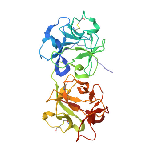
    Viscum album (the European mistletoe) is a semi-parasitic plant, which is of high medical interest. It is widely found in Europe, Asia, and North America. It contains at least three distinct lectins (i.e. ML-I, II, and III), varying in molecular mass and specificity. Among them, ML-I is in focus of medical research for various activities, including anti-cancer activities. To understand the molecular basis for such medical applications, a few studies have already addressed the structural and functional analysis of ML-I in complex with ligands. In continuation of these efforts, we are reporting the crystal structure of ML from Viscum album in complex with the nucleic acid oxidation product 4-N-furfurylcytosine (FC) refined to 2.85 Å resolution. FC is known to be involved in different metabolic pathways related to oxidative stress and DNA modification. X-ray suitable hexagonal crystals of the ML-I/FC complex were grown within four days at 294 K using the hanging drop vapor diffusion method. Diffraction data were collected up to a resolution of 2.85 Å. The ligand affinity was verified by in-silico docking. The high-resolution structure was refined subsequently to analyze particularly the active site conformation and a binding epitope of 4-N-furfurylcytosine. A distinct 2Fo-Fc electron density at the active site was interpreted as a single FC molecule. The specific binding of FC is achieved also through hydrophobic interactions involving Tyr76A, Tyr115A, Glu165A, and Leu157A of the ML-I A-chain. The binding energy of FC to the active site of ML-I was calculated as well to be -6.03 kcal mol-1. In comparison to other reported ML-I complexes, we observed distinct differences in the vicinity of the nucleic acid base binding site upon interaction with FC. Therefore, data obtained will provide new insights in understanding the specificity, inhibition, and cytotoxicity of the ML-I A-chain, and related RIPs.


  • Organizational Affiliation

    Laboratory for Structural Biology of Infection and Inflammation, University of Hamburg, c/o DESY, Build. 22a, Notkestrasse 85, 22607 Hamburg, Germany.


Macromolecules
Find similar proteins by:  (by identity cutoff)  |  3D Structure
Entity ID: 1
MoleculeChains Sequence LengthOrganismDetailsImage
Mistletoe Lectin I249Viscum albumMutation(s): 0 
UniProt
Find proteins for P81446 (Viscum album)
Explore P81446 
Go to UniProtKB:  P81446
Entity Groups  
Sequence Clusters30% Identity50% Identity70% Identity90% Identity95% Identity100% Identity
UniProt GroupP81446
Sequence Annotations
Expand
  • Reference Sequence
Find similar proteins by:  (by identity cutoff)  |  3D Structure
Entity ID: 2
MoleculeChains Sequence LengthOrganismDetailsImage
Mistletoe Lectin I263Viscum albumMutation(s): 0 
UniProt
Find proteins for P81446 (Viscum album)
Explore P81446 
Go to UniProtKB:  P81446
Entity Groups  
Sequence Clusters30% Identity50% Identity70% Identity90% Identity95% Identity100% Identity
UniProt GroupP81446
Sequence Annotations
Expand
  • Reference Sequence
Oligosaccharides

Help

Entity ID: 3
MoleculeChains Length2D Diagram Glycosylation3D Interactions
2-acetamido-2-deoxy-beta-D-glucopyranose-(1-4)-2-acetamido-2-deoxy-beta-D-glucopyranose
C
2N-Glycosylation
Glycosylation Resources
GlyTouCan:  G42666HT
GlyCosmos:  G42666HT
GlyGen:  G42666HT
Entity ID: 4
MoleculeChains Length2D Diagram Glycosylation3D Interactions
alpha-D-galactopyranose-(1-4)-2-acetamido-2-deoxy-beta-D-glucopyranose-(1-4)-2-acetamido-2-deoxy-beta-D-glucopyranose
D
3N-Glycosylation
Glycosylation Resources
GlyTouCan:  G74743ZY
GlyCosmos:  G74743ZY
GlyGen:  G74743ZY
Small Molecules
Ligands 6 Unique
IDChains Name / Formula / InChI Key2D Diagram3D Interactions
NAG
Query on NAG

Download Ideal Coordinates CCD File 
E [auth A],
V [auth B]
2-acetamido-2-deoxy-beta-D-glucopyranose
C8 H15 N O6
OVRNDRQMDRJTHS-FMDGEEDCSA-N
BFW
Query on BFW

Download Ideal Coordinates CCD File 
U [auth A]4-N-Furfurylcytosine
C9 H9 N3 O2
PPZNKEWLJJEHRV-UHFFFAOYSA-N
SO4
Query on SO4

Download Ideal Coordinates CCD File 
K [auth A],
L [auth A],
M [auth A],
N [auth A],
O [auth A]
SULFATE ION
O4 S
QAOWNCQODCNURD-UHFFFAOYSA-L
GOL
Query on GOL

Download Ideal Coordinates CCD File 
AA [auth B]
BA [auth B]
CA [auth B]
DA [auth B]
EA [auth B]
AA [auth B],
BA [auth B],
CA [auth B],
DA [auth B],
EA [auth B],
FA [auth B],
GA [auth B],
HA [auth B],
IA [auth B],
JA [auth B],
KA [auth B],
LA [auth B],
P [auth A],
Q [auth A],
R [auth A],
S [auth A],
Z [auth B]
GLYCEROL
C3 H8 O3
PEDCQBHIVMGVHV-UHFFFAOYSA-N
GLY
Query on GLY

Download Ideal Coordinates CCD File 
MA [auth B],
T [auth A]
GLYCINE
C2 H5 N O2
DHMQDGOQFOQNFH-UHFFFAOYSA-N
CL
Query on CL

Download Ideal Coordinates CCD File 
F [auth A]
G [auth A]
H [auth A]
I [auth A]
J [auth A]
F [auth A],
G [auth A],
H [auth A],
I [auth A],
J [auth A],
W [auth B],
X [auth B],
Y [auth B]
CHLORIDE ION
Cl
VEXZGXHMUGYJMC-UHFFFAOYSA-M
Experimental Data & Validation

Experimental Data

  • Method: X-RAY DIFFRACTION
  • Resolution: 2.84 Å
  • R-Value Free: 0.218 
  • R-Value Work: 0.164 
  • R-Value Observed: 0.166 
  • Space Group: P 65 2 2
Unit Cell:
Length ( Å )Angle ( ˚ )
a = 106.677α = 90
b = 106.677β = 90
c = 312.569γ = 120
Software Package:
Software NamePurpose
REFMACrefinement
XDSdata reduction
SCALAdata scaling
MOLREPphasing

Structure Validation

View Full Validation Report


Ligand Structure Quality Assessment 


Entry History & Funding Information

Deposition Data


Funding OrganizationLocationGrant Number
International Atomic Energy AgencyAustriaINT0086
Hamburg Ministry of Science and ResearchGermanyLFF-GK06

Revision History  (Full details and data files)

  • Version 1.0: 2018-05-02
    Type: Initial release
  • Version 1.1: 2018-07-18
    Changes: Data collection, Database references
  • Version 1.2: 2018-11-28
    Changes: Data collection, Database references
  • Version 1.3: 2019-10-16
    Changes: Data collection
  • Version 2.0: 2020-07-29
    Type: Remediation
    Reason: Carbohydrate remediation
    Changes: Advisory, Atomic model, Data collection, Derived calculations, Structure summary
  • Version 2.1: 2024-01-17
    Changes: Data collection, Database references, Refinement description, Structure summary