


Crystal structure of Type IIP restriction endonuclease PfoI with cognate DNA
Tamulaitiene, G., Manakova, E., Jovaisaite, V., Grazulis, S., Siksnys, V.To be published.
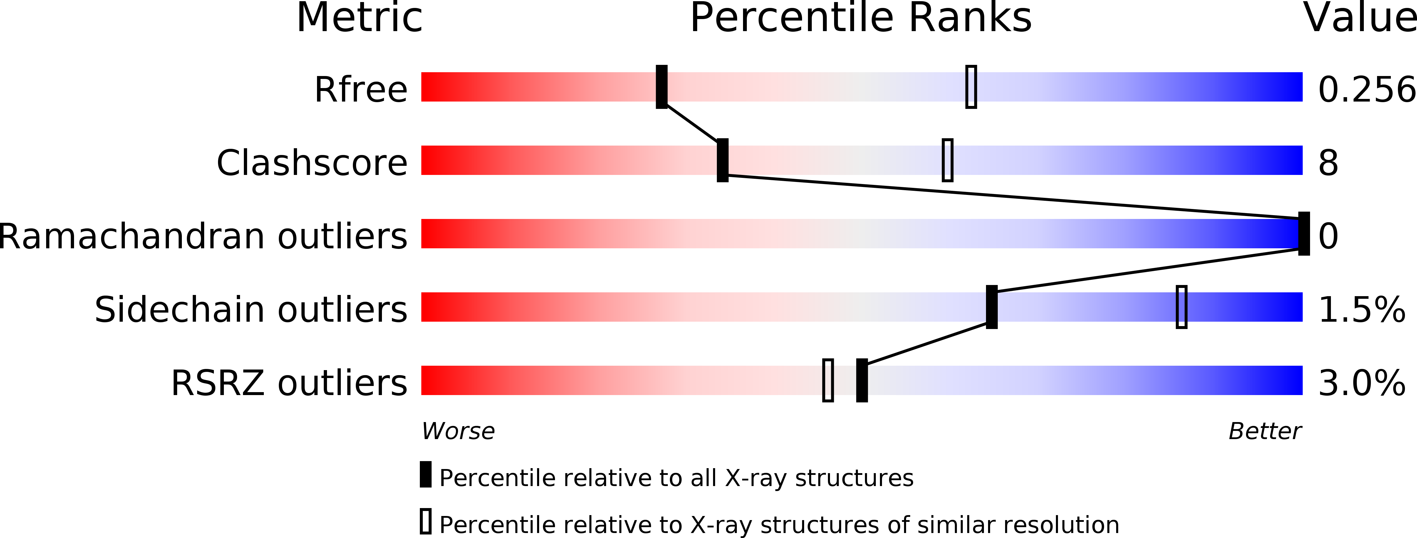
Experimental Data Snapshot
wwPDB Validation 3D Report Full Report
Entity ID: 1 | |||||
|---|---|---|---|---|---|
| Molecule | Chains | Sequence Length | Organism | Details | Image |
| Type ii site-specific deoxyribonuclease | 312 | Klebsiella pneumoniae | Mutation(s): 2 EC: 3.1.21.4 | 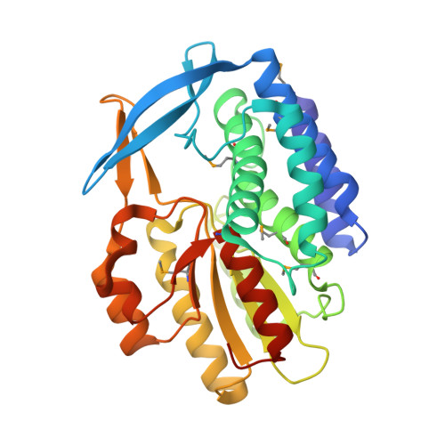 | |
UniProt | |||||
Find proteins for Q93K38 (Klebsiella pneumoniae) Explore Q93K38 Go to UniProtKB: Q93K38 | |||||
Entity Groups | |||||
| Sequence Clusters | 30% Identity50% Identity70% Identity90% Identity95% Identity100% Identity | ||||
| UniProt Group | Q93K38 | ||||
Sequence AnnotationsExpand | |||||
| |||||
| Ligands 1 Unique | |||||
|---|---|---|---|---|---|
| ID | Chains | Name / Formula / InChI Key | 2D Diagram | 3D Interactions | |
| SO4 Query on SO4 | B [auth A], C [auth A], D [auth A], E [auth A], F [auth A] | SULFATE ION O4 S QAOWNCQODCNURD-UHFFFAOYSA-L |  | ||
| Modified Residues 1 Unique | |||||
|---|---|---|---|---|---|
| ID | Chains | Type | Formula | 2D Diagram | Parent |
| MSE Query on MSE | A | L-PEPTIDE LINKING | C5 H11 N O2 Se |  | MET |
| Length ( Å ) | Angle ( ˚ ) |
|---|---|
| a = 134.794 | α = 90 |
| b = 134.794 | β = 90 |
| c = 134.794 | γ = 90 |
| Software Name | Purpose |
|---|---|
| REFMAC | refinement |
| XDS | data reduction |
| SCALA | data scaling |
| Auto-Rickshaw | phasing |
| Funding Organization | Location | Grant Number |
|---|---|---|
| Research Council of Lithuania | Lithuania | MIP-41/2013 |
| European Union | BioStruct-X (grant agreement 283570) |
RCSB PDB (citation) is hosted by
RCSB PDB is a member of the

