6EEY

Crystal structure of human Scribble PDZ4 R1110G Mutant


Experimental Data Snapshot

  • Method: X-RAY DIFFRACTION
  • Resolution: 1.15 Å
  • R-Value Free: 0.182 
  • R-Value Work: 0.157 
  • R-Value Observed: 0.160 

wwPDB Validation   3D Report Full Report


This is version 1.4 of the entry. See complete history


Literature

Scribble co-operatively binds multiple alpha1D-adrenergic receptor C-terminal PDZ ligands.

Janezic, E.M.Harris, D.A.Dinh, D.Lee, K.S.Stewart, A.Hinds, T.R.Hsu, P.L.Zheng, N.Hague, C.

(2019) Sci Rep 9: 14073-14073

  • DOI: https://doi.org/10.1038/s41598-019-50671-6
  • Primary Citation of Related Structures:  
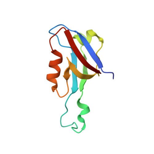
    6EEY

  • PubMed Abstract: 

    Many G protein-coupled receptors (GPCRs) are organized as dynamic macromolecular complexes in human cells. Unraveling the structural determinants of unique GPCR complexes may identify unique protein:protein interfaces to be exploited for drug development. We previously reported α 1D -adrenergic receptors (α 1D -ARs) - key regulators of cardiovascular and central nervous system function - form homodimeric, modular PDZ protein complexes with cell-type specificity. Towards mapping α 1D -AR complex architecture, biolayer interferometry (BLI) revealed the α 1D -AR C-terminal PDZ ligand selectively binds the PDZ protein scribble (SCRIB) with >8x higher affinity than known interactors syntrophin, CASK and DLG1. Complementary in situ and in vitro assays revealed SCRIB PDZ domains 1 and 4 to be high affinity α 1D -AR PDZ ligand interaction sites. SNAP-GST pull-down assays demonstrate SCRIB binds multiple α 1D -AR PDZ ligands via a co-operative mechanism. Structure-function analyses pinpoint R1110 PDZ4 as a unique, critical residue dictating SCRIB:α 1D -AR binding specificity. The crystal structure of SCRIB PDZ4 R1110G predicts spatial shifts in the SCRIB PDZ4 carboxylate binding loop dictate α 1D -AR binding specificity. Thus, the findings herein identify SCRIB PDZ domains 1 and 4 as high affinity α 1D -AR interaction sites, and potential drug targets to treat diseases associated with aberrant α 1D -AR signaling.


  • Organizational Affiliation

    Department of Pharmacology, School of Medicine, University of Washington, 1959 NE Pacific Street, Seattle, WA, 98195, USA.


Macromolecules
Find similar proteins by:  (by identity cutoff)  |  3D Structure
Entity ID: 1
MoleculeChains Sequence LengthOrganismDetailsImage
Protein scribble homolog95Homo sapiensMutation(s): 1 
Gene Names: SCRIBCRIB1KIAA0147LAP4SCRB1VARTUL
UniProt & NIH Common Fund Data Resources
Find proteins for Q14160 (Homo sapiens)
Explore Q14160 
Go to UniProtKB:  Q14160
PHAROS:  Q14160
GTEx:  ENSG00000180900 
Entity Groups  
Sequence Clusters30% Identity50% Identity70% Identity90% Identity95% Identity100% Identity
UniProt GroupQ14160
Sequence Annotations
Expand
  • Reference Sequence
Experimental Data & Validation

Experimental Data

  • Method: X-RAY DIFFRACTION
  • Resolution: 1.15 Å
  • R-Value Free: 0.182 
  • R-Value Work: 0.157 
  • R-Value Observed: 0.160 
  • Space Group: P 1 21 1
Unit Cell:
Length ( Å )Angle ( ˚ )
a = 27.286α = 90
b = 40.235β = 97.85
c = 32.26γ = 90
Software Package:
Software NamePurpose
Aimlessdata scaling
PHASERphasing
PHENIXrefinement
PDB_EXTRACTdata extraction
XDSdata reduction

Structure Validation

View Full Validation Report


Entry History & Funding Information

Deposition Data


Funding OrganizationLocationGrant Number
National Institutes of Health/National Institute of General Medical Sciences (NIH/NIGMS)United StatesT32GM007750
National Institutes of Health/National Institute of General Medical Sciences (NIH/NIGMS)United StatesR01GM100893

Revision History  (Full details and data files)

  • Version 1.0: 2019-10-02
    Type: Initial release
  • Version 1.1: 2019-10-09
    Changes: Data collection, Database references
  • Version 1.2: 2019-10-16
    Changes: Data collection, Database references
  • Version 1.3: 2020-01-01
    Changes: Author supporting evidence
  • Version 1.4: 2023-10-11
    Changes: Data collection, Database references, Refinement description