6DS2

Crystal structure of Ni(II)-bound human calprotectin


Experimental Data Snapshot

  • Method: X-RAY DIFFRACTION
  • Resolution: 2.10 Å
  • R-Value Free: 0.222 
  • R-Value Work: 0.185 
  • R-Value Observed: 0.187 

wwPDB Validation   3D Report Full Report


This is version 1.3 of the entry. See complete history


Literature

Biophysical Examination of the Calcium-Modulated Nickel-Binding Properties of Human Calprotectin Reveals Conformational Change in the EF-Hand Domains and His3Asp Site.

Nakashige, T.G.Bowman, S.E.J.Zygiel, E.M.Drennan, C.L.Nolan, E.M.

(2018) Biochemistry 57: 4155-4164

  • DOI: https://doi.org/10.1021/acs.biochem.8b00415
  • Primary Citation of Related Structures:  
    6DS2

  • PubMed Abstract: 

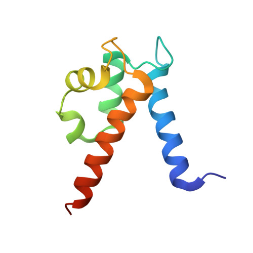
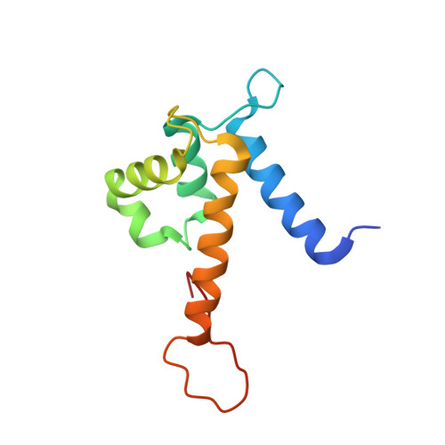
    Calprotectin (CP, S100A8/S100A9 oligomer, MRP-8/MRP-14 oligomer) is a host-defense protein that sequesters nutrient transition metals from microbes. Each S100A8/S100A9 heterodimer contains four EF-hand domains and two transition-metal-binding sites. We investigate the effect of Ca(II) ions on the structure and Ni(II)-binding properties of human CP. By employing energy dispersive X-ray (EDX) spectroscopy, we evaluate the metal content of Ni(II)-bound CP-Ser [oligomer of S100A8(C42S) and S100A9(C3S)] crystals obtained in the absence and presence of Ca(II). We present a 2.1 Å resolution crystal structure of Ni(II)-bound CP-Ser and compare this structure to a reported Ni(II)- and Ca(II)-bound CP-Ser structure [Nakashige, T. G., et al. (2017) J. Am. Chem. Soc. 139, 8828-8836]. This analysis reveals conformational changes associated with coordination of Ca(II) to the EF-hands of S100A9 and that Ca(II) binding affects the coordination number and geometry of the Ni(II) ion bound to the His 3 Asp site. In contrast, negligible differences are observed for the Ni(II)-His 6 site in the absence and presence of Ca(II). Biochemical studies show that, whereas the His 6 site has a thermodynamic preference for Ni(II) over Zn(II), the His 3 Asp site selects for Zn(II) over Ni(II), and relatively rapid metal exchange occurs at this site. These observations inform the working model for how CP withholds nutrient metals in the extracellular space.


Macromolecules
Find similar proteins by:  (by identity cutoff)  |  3D Structure
Entity ID: 1
MoleculeChains Sequence LengthOrganismDetailsImage
Protein S100-A8
A, C, E, G
93Homo sapiensMutation(s): 1 
Gene Names: S100A8CAGACFAGMRP8
UniProt & NIH Common Fund Data Resources
Find proteins for P05109 (Homo sapiens)
Explore P05109 
Go to UniProtKB:  P05109
PHAROS:  P05109
GTEx:  ENSG00000143546 
Entity Groups  
Sequence Clusters30% Identity50% Identity70% Identity90% Identity95% Identity100% Identity
UniProt GroupP05109
Sequence Annotations
Expand
  • Reference Sequence
Find similar proteins by:  (by identity cutoff)  |  3D Structure
Entity ID: 2
MoleculeChains Sequence LengthOrganismDetailsImage
Protein S100-A9
B, D, F, H
114Homo sapiensMutation(s): 0 
Gene Names: S100A9CAGBCFAGMRP14
UniProt & NIH Common Fund Data Resources
Find proteins for P06702 (Homo sapiens)
Explore P06702 
Go to UniProtKB:  P06702
PHAROS:  P06702
GTEx:  ENSG00000163220 
Entity Groups  
Sequence Clusters30% Identity50% Identity70% Identity90% Identity95% Identity100% Identity
UniProt GroupP06702
Sequence Annotations
Expand
  • Reference Sequence
Small Molecules
Ligands 2 Unique
IDChains Name / Formula / InChI Key2D Diagram3D Interactions
NI (Subject of Investigation/LOI)
Query on NI

Download Ideal Coordinates CCD File 
K [auth B]
L [auth C]
O [auth D]
Q [auth E]
T [auth F]
K [auth B],
L [auth C],
O [auth D],
Q [auth E],
T [auth F],
X [auth H]
NICKEL (II) ION
Ni
VEQPNABPJHWNSG-UHFFFAOYSA-N
NA
Query on NA

Download Ideal Coordinates CCD File 
I [auth A]
J [auth A]
M [auth C]
N [auth C]
P [auth D]
I [auth A],
J [auth A],
M [auth C],
N [auth C],
P [auth D],
R [auth E],
S [auth E],
U [auth F],
V [auth G],
W [auth G]
SODIUM ION
Na
FKNQFGJONOIPTF-UHFFFAOYSA-N
Experimental Data & Validation

Experimental Data

  • Method: X-RAY DIFFRACTION
  • Resolution: 2.10 Å
  • R-Value Free: 0.222 
  • R-Value Work: 0.185 
  • R-Value Observed: 0.187 
  • Space Group: P 21 21 21
Unit Cell:
Length ( Å )Angle ( ˚ )
a = 57.145α = 90
b = 78.082β = 90
c = 223.787γ = 90
Software Package:
Software NamePurpose
PHENIXrefinement
HKL-2000data scaling
HKL-2000data reduction
PHENIXphasing

Structure Validation

View Full Validation Report


Entry History & Funding Information

Deposition Data


Funding OrganizationLocationGrant Number
National Science Foundation (NSF, United States)United StatesCHE-1352132
National Institutes of Health/National Institute of General Medical Sciences (NIH/NIGMS)United StatesR01GM126376
National Institutes of Health/National Institute of General Medical Sciences (NIH/NIGMS)United StatesR01GM118695
National Institutes of Health/National Institute of General Medical Sciences (NIH/NIGMS)United StatesR01GM069857

Revision History  (Full details and data files)

  • Version 1.0: 2018-07-04
    Type: Initial release
  • Version 1.1: 2018-08-01
    Changes: Data collection, Database references
  • Version 1.2: 2019-11-27
    Changes: Author supporting evidence
  • Version 1.3: 2023-10-11
    Changes: Data collection, Database references, Derived calculations, Refinement description