6DDU

mouse beta-mannosidase bound to beta-D-mannose (MANBA)


Experimental Data Snapshot

  • Method: X-RAY DIFFRACTION
  • Resolution: 2.67 Å
  • R-Value Free: 0.246 
  • R-Value Work: 0.199 
  • R-Value Observed: 0.203 

wwPDB Validation   3D Report Full Report


Ligand Structure Quality Assessment 


This is version 2.1 of the entry. See complete history


Literature

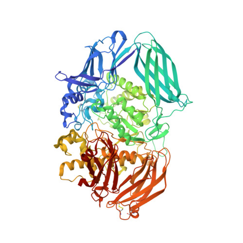
The structure of mammalian beta-mannosidase provides insight into beta-mannosidosis and nystagmus.

Gytz, H.Liang, J.Liang, Y.Gorelik, A.Illes, K.Nagar, B.

(2019) FEBS J 286: 1319-1331

  • DOI: https://doi.org/10.1111/febs.14731
  • Primary Citation of Related Structures:  
    6DDT, 6DDU

  • PubMed Abstract: 

    β-Mannosidase is a lysosomal enzyme from the glycosyl hydrolase family 2 that cleaves the single β(1-4)-linked mannose at the nonreducing end of N-glycosylated proteins, and plays an important role in the polysaccharide degradation pathway. Mutations in the MANBA gene, which encodes the β-mannosidase, can lead to the lysosomal storage disease β-mannosidosis, as well as nystagmus, an eye condition characterized by involuntary eye movements. Here, we present the first structures of a mammalian β-mannosidase in both the apo- and mannose-bound forms. The structure is similar to previously determined β-mannosidase structures with regard to domain organization and fold, however, there are important differences that underlie substrate specificity between species. Additionally, in contrast to most other ligand-bound β-mannosidases from bacterial and fungal sources where bound sugars were in a boat-like conformation, we find the mannose in the chair conformation. Evaluation of known disease mutations in the MANBA gene provides insight into their impact on disease phenotypes. Together, these results will be important for the design of therapeutics for treating diseases caused by β-mannosidase deficiency. DATABASE: Structural data are available in the Protein Data Bank under the accession numbers 6DDT and 6DDU.


  • Organizational Affiliation

    Department of Biochemistry, Groupe de Recherche Axé sur la Structure des Protéines, McGill University, Montreal, Canada.


Macromolecules
Find similar proteins by:  (by identity cutoff)  |  3D Structure
Entity ID: 1
MoleculeChains Sequence LengthOrganismDetailsImage
Beta-mannosidase868Mus musculusMutation(s): 0 
Gene Names: ManbaBmn
EC: 3.2.1.25
UniProt
Find proteins for Q8K2I4 (Mus musculus)
Explore Q8K2I4 
Go to UniProtKB:  Q8K2I4
Entity Groups  
Sequence Clusters30% Identity50% Identity70% Identity90% Identity95% Identity100% Identity
UniProt GroupQ8K2I4
Sequence Annotations
Expand
  • Reference Sequence
Oligosaccharides

Help

Entity ID: 2
MoleculeChains Length2D Diagram Glycosylation3D Interactions
alpha-L-fucopyranose-(1-6)-2-acetamido-2-deoxy-beta-D-glucopyranose
B, D
2N-Glycosylation
Glycosylation Resources
GlyTouCan:  G86851RC
GlyCosmos:  G86851RC
GlyGen:  G86851RC
Entity ID: 3
MoleculeChains Length2D Diagram Glycosylation3D Interactions
2-acetamido-2-deoxy-beta-D-glucopyranose-(1-4)-2-acetamido-2-deoxy-beta-D-glucopyranose
C
2N-Glycosylation
Glycosylation Resources
GlyTouCan:  G42666HT
GlyCosmos:  G42666HT
GlyGen:  G42666HT
Small Molecules
Ligands 4 Unique
IDChains Name / Formula / InChI Key2D Diagram3D Interactions
NAG
Query on NAG

Download Ideal Coordinates CCD File 
F [auth A],
G [auth A],
H [auth A]
2-acetamido-2-deoxy-beta-D-glucopyranose
C8 H15 N O6
OVRNDRQMDRJTHS-FMDGEEDCSA-N
BMA (Subject of Investigation/LOI)
Query on BMA

Download Ideal Coordinates CCD File 
I [auth A]beta-D-mannopyranose
C6 H12 O6
WQZGKKKJIJFFOK-RWOPYEJCSA-N
EDO
Query on EDO

Download Ideal Coordinates CCD File 
J [auth A]1,2-ETHANEDIOL
C2 H6 O2
LYCAIKOWRPUZTN-UHFFFAOYSA-N
CA
Query on CA

Download Ideal Coordinates CCD File 
E [auth A]CALCIUM ION
Ca
BHPQYMZQTOCNFJ-UHFFFAOYSA-N
Experimental Data & Validation

Experimental Data

  • Method: X-RAY DIFFRACTION
  • Resolution: 2.67 Å
  • R-Value Free: 0.246 
  • R-Value Work: 0.199 
  • R-Value Observed: 0.203 
  • Space Group: P 1 21 1
Unit Cell:
Length ( Å )Angle ( ˚ )
a = 81.162α = 90
b = 65.651β = 92.73
c = 104.531γ = 90
Software Package:
Software NamePurpose
PHENIXrefinement
HKL-2000data reduction
HKL-2000data scaling
PHASERphasing

Structure Validation

View Full Validation Report


Ligand Structure Quality Assessment 


Entry History & Funding Information

Deposition Data


Funding OrganizationLocationGrant Number
Canadian Institutes of Health Research (CIHR)CanadaMOP-133535

Revision History  (Full details and data files)

  • Version 1.0: 2019-01-09
    Type: Initial release
  • Version 1.1: 2019-04-17
    Changes: Data collection, Database references
  • Version 1.2: 2020-01-08
    Changes: Author supporting evidence, Data collection
  • Version 2.0: 2020-07-29
    Type: Remediation
    Reason: Carbohydrate remediation
    Changes: Atomic model, Data collection, Derived calculations, Structure summary
  • Version 2.1: 2023-10-11
    Changes: Data collection, Database references, Derived calculations, Refinement description, Structure summary