6AZV

IDO1/BMS-978587 crystal structure


Experimental Data Snapshot

  • Method: X-RAY DIFFRACTION
  • Resolution: 2.75 Å
  • R-Value Free: 0.272 
  • R-Value Work: 0.195 
  • R-Value Observed: 0.199 

wwPDB Validation   3D Report Full Report


Ligand Structure Quality Assessment 


This is version 1.2 of the entry. See complete history


Literature

Immune-modulating enzyme indoleamine 2,3-dioxygenase is effectively inhibited by targeting its apo-form.

Nelp, M.T.Kates, P.A.Hunt, J.T.Newitt, J.A.Balog, A.Maley, D.Zhu, X.Abell, L.Allentoff, A.Borzilleri, R.Lewis, H.A.Lin, Z.Seitz, S.P.Yan, C.Groves, J.T.

(2018) Proc Natl Acad Sci U S A 115: 3249-3254

  • DOI: https://doi.org/10.1073/pnas.1719190115
  • Primary Citation of Related Structures:  
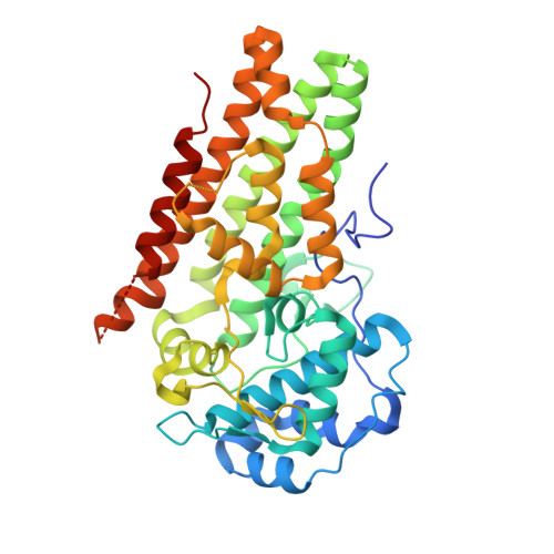
    6AZU, 6AZV, 6AZW

  • PubMed Abstract: 

    For cancer cells to survive and proliferate, they must escape normal immune destruction. One mechanism by which this is accomplished is through immune suppression effected by up-regulation of indoleamine 2,3-dioxygenase (IDO1), a heme enzyme that catalyzes the oxidation of tryptophan to N -formylkynurenine. On deformylation, kynurenine and downstream metabolites suppress T cell function. The importance of this immunosuppressive mechanism has spurred intense interest in the development of clinical IDO1 inhibitors. Herein, we describe the mechanism by which a class of compounds effectively and specifically inhibits IDO1 by targeting its apo-form. We show that the in vitro kinetics of inhibition coincide with an unusually high rate of intrinsic enzyme-heme dissociation, especially in the ferric form. X-ray crystal structures of the inhibitor-enzyme complexes show that heme is displaced from the enzyme and blocked from rebinding by these compounds. The results reveal that apo-IDO1 serves as a unique target for inhibition and that heme lability plays an important role in posttranslational regulation.


  • Organizational Affiliation

    Department of Chemistry, Princeton University, Princeton, NJ 08544.


Macromolecules
Find similar proteins by:  (by identity cutoff)  |  3D Structure
Entity ID: 1
MoleculeChains Sequence LengthOrganismDetailsImage
Indoleamine 2,3-dioxygenase 1
A, B, C, D
402Homo sapiensMutation(s): 0 
Gene Names: IDO1IDOINDO
EC: 1.13.11.52
UniProt & NIH Common Fund Data Resources
Find proteins for P14902 (Homo sapiens)
Explore P14902 
Go to UniProtKB:  P14902
PHAROS:  P14902
GTEx:  ENSG00000131203 
Entity Groups  
Sequence Clusters30% Identity50% Identity70% Identity90% Identity95% Identity100% Identity
UniProt GroupP14902
Sequence Annotations
Expand
  • Reference Sequence
Experimental Data & Validation

Experimental Data

  • Method: X-RAY DIFFRACTION
  • Resolution: 2.75 Å
  • R-Value Free: 0.272 
  • R-Value Work: 0.195 
  • R-Value Observed: 0.199 
  • Space Group: C 1 2 1
Unit Cell:
Length ( Å )Angle ( ˚ )
a = 203.392α = 90
b = 120.868β = 119.11
c = 101.527γ = 90
Software Package:
Software NamePurpose
SCALAdata scaling
PHENIXrefinement
PDB_EXTRACTdata extraction
XDSdata reduction
PHENIXphasing

Structure Validation

View Full Validation Report


Ligand Structure Quality Assessment 


Entry History 

Deposition Data

  • Released Date: 2018-03-21 
  • Deposition Author(s): Lewis, H.A.

Revision History  (Full details and data files)

  • Version 1.0: 2018-03-21
    Type: Initial release
  • Version 1.1: 2018-04-11
    Changes: Data collection, Database references
  • Version 1.2: 2023-10-04
    Changes: Data collection, Database references, Refinement description