6AX7

The crystal structure of a lysyl hydroxylase from Acanthamoeba polyphaga mimivirus


Experimental Data Snapshot

  • Method: X-RAY DIFFRACTION
  • Resolution: 2.00 Å
  • R-Value Free: 0.216 
  • R-Value Work: 0.183 
  • R-Value Observed: 0.184 

wwPDB Validation   3D Report Full Report


This is version 1.3 of the entry. See complete history


Literature

Pro-metastatic collagen lysyl hydroxylase dimer assemblies stabilized by Fe2+-binding.

Guo, H.F.Tsai, C.L.Terajima, M.Tan, X.Banerjee, P.Miller, M.D.Liu, X.Yu, J.Byemerwa, J.Alvarado, S.Kaoud, T.S.Dalby, K.N.Bota-Rabassedas, N.Chen, Y.Yamauchi, M.Tainer, J.A.Phillips, G.N.Kurie, J.M.

(2018) Nat Commun 9: 512-512

  • DOI: https://doi.org/10.1038/s41467-018-02859-z
  • Primary Citation of Related Structures:  
    6AX6, 6AX7

  • PubMed Abstract: 

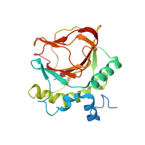
    Collagen lysyl hydroxylases (LH1-3) are Fe 2+ - and 2-oxoglutarate (2-OG)-dependent oxygenases that maintain extracellular matrix homeostasis. High LH2 levels cause stable collagen cross-link accumulations that promote fibrosis and cancer progression. However, developing LH antagonists will require structural insights. Here, we report a 2 Å crystal structure and X-ray scattering on dimer assemblies for the LH domain of L230 in Acanthamoeba polyphaga mimivirus. Loop residues in the double-stranded β-helix core generate a tail-to-tail dimer. A stabilizing hydrophobic leucine locks into an aromatic tyrosine-pocket on the opposite subunit. An active site triad coordinates Fe 2+ . The two active sites flank a deep surface cleft that suggest dimerization creates a collagen-binding site. Loss of Fe 2+ -binding disrupts the dimer. Dimer disruption and charge reversal in the cleft increase K m and reduce LH activity. Ectopic L230 expression in tumors promotes collagen cross-linking and metastasis. These insights suggest inhibitor targets for fibrosis and cancer.


  • Organizational Affiliation

    Department of Thoracic/Head and Neck Medical Oncology, The University of Texas MD Anderson Cancer Center, Houston, TX, 77030, USA.


Macromolecules
Find similar proteins by:  (by identity cutoff)  |  3D Structure
Entity ID: 1
MoleculeChains Sequence LengthOrganismDetailsImage
Procollagen lysyl hydroxylase and glycosyltransferase
A, B
235Acanthamoeba polyphaga mimivirusMutation(s): 0 
EC: 1.14.11.4
UniProt
Find proteins for Q5UQC3 (Acanthamoeba polyphaga mimivirus)
Explore Q5UQC3 
Go to UniProtKB:  Q5UQC3
Entity Groups  
Sequence Clusters30% Identity50% Identity70% Identity90% Identity95% Identity100% Identity
UniProt GroupQ5UQC3
Sequence Annotations
Expand
  • Reference Sequence
Experimental Data & Validation

Experimental Data

  • Method: X-RAY DIFFRACTION
  • Resolution: 2.00 Å
  • R-Value Free: 0.216 
  • R-Value Work: 0.183 
  • R-Value Observed: 0.184 
  • Space Group: P 43 21 2
Unit Cell:
Length ( Å )Angle ( ˚ )
a = 109.46α = 90
b = 109.46β = 90
c = 84.023γ = 90
Software Package:
Software NamePurpose
PHENIXrefinement
XDSdata reduction
XDSdata scaling
PHENIXphasing

Structure Validation

View Full Validation Report


Entry History & Funding Information

Deposition Data


Funding OrganizationLocationGrant Number
National Institutes of Health/National Cancer Institute (NIH/NCI)United StatesR01CA105155
National Institutes of Health/National Cancer Institute (NIH/NCI)United StatesP50CA70907
Cancer Prevention and Research Institute of Texas (CPRIT)United StatesRP160652

Revision History  (Full details and data files)

  • Version 1.0: 2018-02-21
    Type: Initial release
  • Version 1.1: 2018-02-28
    Changes: Author supporting evidence
  • Version 1.2: 2019-12-04
    Changes: Author supporting evidence
  • Version 1.3: 2023-10-04
    Changes: Data collection, Database references, Derived calculations, Refinement description