


Crystal structure of Nucleoside diphosphate kinase from Pseudomonas aeruginosa at 3.55 A resolution.
Sikarwar, J., Singh, P.K., Sharma, S., Singh, T.P.To be published.
Experimental Data Snapshot
Starting Model: experimental
View more details
wwPDB Validation 3D Report Full Report

Entity ID: 1 | |||||
|---|---|---|---|---|---|
| Molecule | Chains | Sequence Length | Organism | Details | Image |
| Nucleoside diphosphate kinase | 143 | Pseudomonas aeruginosa | Mutation(s): 0 EC: 2.7.4.6 | 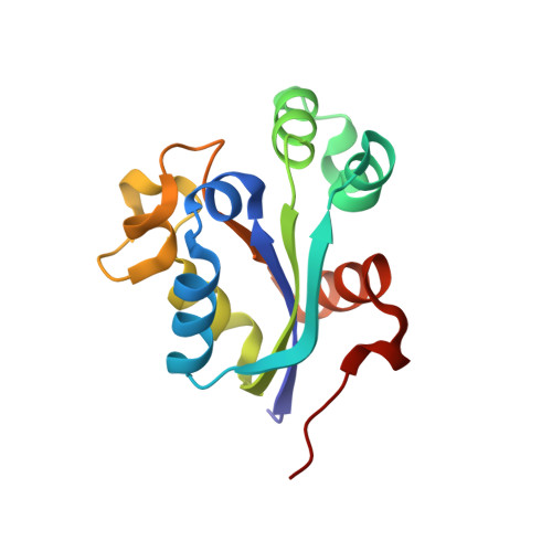 | |
Entity Groups | |||||
| Sequence Clusters | 30% Identity50% Identity70% Identity90% Identity95% Identity100% Identity | ||||
Sequence AnnotationsExpand | |||||
| |||||
| Length ( Å ) | Angle ( ˚ ) |
|---|---|
| a = 68.566 | α = 99.6 |
| b = 70.875 | β = 109.12 |
| c = 71.097 | γ = 90.25 |
| Software Name | Purpose |
|---|---|
| REFMAC | refinement |
| XDS | data reduction |
| autoPROC | data scaling |
| MOLREP | phasing |
RCSB PDB (citation) is hosted by
RCSB PDB is a member of the

