5ZRZ

Crystal Structure of EphA5/SAMD5 Complex


Experimental Data Snapshot

  • Method: X-RAY DIFFRACTION
  • Resolution: 1.89 Å
  • R-Value Free: 0.234 
  • R-Value Work: 0.194 
  • R-Value Observed: 0.196 

wwPDB Validation   3D Report Full Report


This is version 1.1 of the entry. See complete history


Literature

Specific Eph receptor-cytoplasmic effector signaling mediated by SAM-SAM domain interactions.

Wang, Y.Shang, Y.Li, J.Chen, W.Li, G.Wan, J.Liu, W.Zhang, M.

(2018) Elife 7

  • DOI: https://doi.org/10.7554/eLife.35677
  • Primary Citation of Related Structures:  
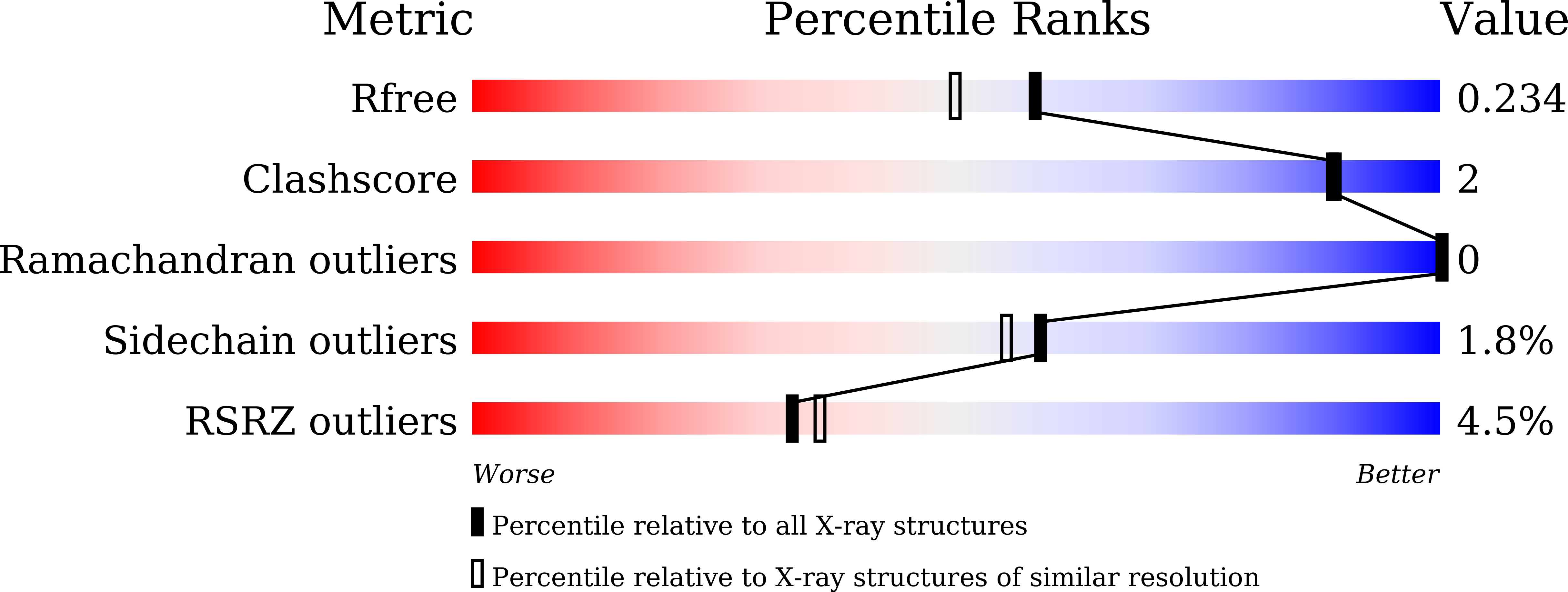
    5ZRX, 5ZRY, 5ZRZ

  • PubMed Abstract: 

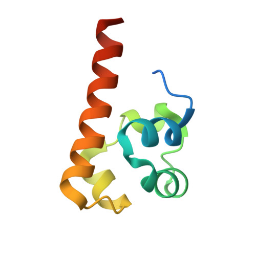
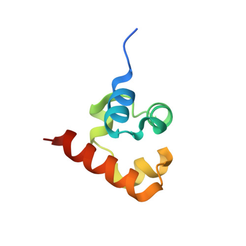
    The Eph receptor tyrosine kinase (RTK) family is the largest subfamily of RTKs playing critical roles in many developmental processes such as tissue patterning, neurogenesis and neuronal circuit formation, angiogenesis, etc. How the 14 Eph proteins, via their highly similar cytoplasmic domains, can transmit diverse and sometimes opposite cellular signals upon engaging ephrins is a major unresolved question. Here, we systematically investigated the bindings of each SAM domain of Eph receptors to the SAM domains from SHIP2 and Odin, and uncover a highly specific SAM-SAM interaction-mediated cytoplasmic Eph-effector binding pattern. Comparative X-ray crystallographic studies of several SAM-SAM heterodimer complexes, together with biochemical and cell biology experiments, not only revealed the exquisite specificity code governing Eph/effector interactions but also allowed us to identify SAMD5 as a new Eph binding partner. Finally, these Eph/effector SAM heterodimer structures can explain many Eph SAM mutations identified in patients suffering from cancers and other diseases.


  • Organizational Affiliation

    Shenzhen Key Laboratory for Neuronal Structural Biology, Biomedical Research Institute, Shenzhen Peking University-The Hong Kong University of Science and Technology Medical Center, Shenzhen, China.


Macromolecules
Find similar proteins by:  (by identity cutoff)  |  3D Structure
Entity ID: 1
MoleculeChains Sequence LengthOrganismDetailsImage
Ephrin type-A receptor 580Mus musculusMutation(s): 0 
Gene Names: Epha5BskEhk1
EC: 2.7.10.1
UniProt & NIH Common Fund Data Resources
Find proteins for Q60629 (Mus musculus)
Explore Q60629 
Go to UniProtKB:  Q60629
IMPC:  MGI:99654
Entity Groups  
Sequence Clusters30% Identity50% Identity70% Identity90% Identity95% Identity100% Identity
UniProt GroupQ60629
Sequence Annotations
Expand
  • Reference Sequence
Find similar proteins by:  (by identity cutoff)  |  3D Structure
Entity ID: 2
MoleculeChains Sequence LengthOrganismDetailsImage
Sterile alpha motif domain-containing protein 570Mus musculusMutation(s): 0 
Gene Names: Samd5E130306M17RikmCG_52174
UniProt
Find proteins for Q3V1H9 (Mus musculus)
Explore Q3V1H9 
Go to UniProtKB:  Q3V1H9
Entity Groups  
Sequence Clusters30% Identity50% Identity70% Identity90% Identity95% Identity100% Identity
UniProt GroupQ3V1H9
Sequence Annotations
Expand
  • Reference Sequence
Experimental Data & Validation

Experimental Data

  • Method: X-RAY DIFFRACTION
  • Resolution: 1.89 Å
  • R-Value Free: 0.234 
  • R-Value Work: 0.194 
  • R-Value Observed: 0.196 
  • Space Group: C 1 2 1
Unit Cell:
Length ( Å )Angle ( ˚ )
a = 98.195α = 90
b = 29.651β = 109.95
c = 54.17γ = 90
Software Package:
Software NamePurpose
DENZOdata reduction
SCALEPACKdata scaling
REFMACrefinement
PDB_EXTRACTdata extraction
PHASERphasing

Structure Validation

View Full Validation Report


Entry History & Funding Information

Deposition Data


Funding OrganizationLocationGrant Number
National Key R&D Program of ChinaChina2016YFA0501903

Revision History  (Full details and data files)

  • Version 1.0: 2018-05-30
    Type: Initial release
  • Version 1.1: 2023-11-22
    Changes: Data collection, Database references, Refinement description