5ZHP

M3 muscarinic acetylcholine receptor in complex with a selective antagonist


Experimental Data Snapshot

  • Method: X-RAY DIFFRACTION
  • Resolution: 3.10 Å
  • R-Value Free: 0.267 
  • R-Value Work: 0.229 
  • R-Value Observed: 0.232 

wwPDB Validation   3D Report Full Report


Ligand Structure Quality Assessment 


This is version 2.1 of the entry. See complete history


Literature

Structure-guided development of selective M3 muscarinic acetylcholine receptor antagonists

Liu, H.Hofmann, J.Fish, I.Schaake, B.Eitel, K.Bartuschat, A.Kaindl, J.Rampp, H.Banerjee, A.Hubner, H.Clark, M.J.Vincent, S.G.Fisher, J.T.Heinrich, M.R.Hirata, K.Liu, X.Sunahara, R.K.Shoichet, B.K.Kobilka, B.K.Gmeiner, P.

(2018) Proc Natl Acad Sci U S A 115: 12046-12050

  • DOI: https://doi.org/10.1073/pnas.1813988115
  • Primary Citation of Related Structures:  
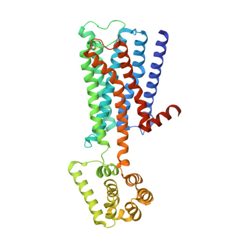
    5ZHP

  • PubMed Abstract: 

    Drugs that treat chronic obstructive pulmonary disease by antagonizing the M3 muscarinic acetylcholine receptor (M3R) have had a significant effect on health, but can suffer from their lack of selectivity against the M2R subtype, which modulates heart rate. Beginning with the crystal structures of M2R and M3R, we exploited a single amino acid difference in their orthosteric binding pockets using molecular docking and structure-based design. The resulting M3R antagonists had up to 100-fold selectivity over M2R in affinity and over 1,000-fold selectivity in vivo. The crystal structure of the M3R-selective antagonist in complex with M3R corresponded closely to the docking-predicted geometry, providing a template for further optimization.


  • Organizational Affiliation

    Beijing Advanced Innovation Center for Structural Biology, School of Medicine, Tsinghua University, 100084 Beijing, China.


Macromolecules
Find similar proteins by:  (by identity cutoff)  |  3D Structure
Entity ID: 1
MoleculeChains Sequence LengthOrganismDetailsImage
Muscarinic acetylcholine receptor M3,Endolysin,Endolysin,Muscarinic acetylcholine receptor M3
A, B
418Rattus norvegicusTequatrovirus T4Mutation(s): 2 
Gene Names: Chrm3Chrm-3e
EC: 3.2.1.17
UniProt
Find proteins for P08483 (Rattus norvegicus)
Explore P08483 
Go to UniProtKB:  P08483
Find proteins for P00720 (Enterobacteria phage T4)
Explore P00720 
Go to UniProtKB:  P00720
Entity Groups  
Sequence Clusters30% Identity50% Identity70% Identity90% Identity95% Identity100% Identity
UniProt GroupsP00720P08483
Sequence Annotations
Expand
  • Reference Sequence
Oligosaccharides

Help

Entity ID: 2
MoleculeChains Length2D Diagram Glycosylation3D Interactions
alpha-D-glucopyranose-(1-4)-alpha-D-glucopyranose
C
2N/A
Glycosylation Resources
GlyTouCan:  G07411ON
GlyCosmos:  G07411ON
Small Molecules
Biologically Interesting Molecules (External Reference) 1 Unique
Experimental Data & Validation

Experimental Data

  • Method: X-RAY DIFFRACTION
  • Resolution: 3.10 Å
  • R-Value Free: 0.267 
  • R-Value Work: 0.229 
  • R-Value Observed: 0.232 
  • Space Group: P 1 2 1
Unit Cell:
Length ( Å )Angle ( ˚ )
a = 122.11α = 90
b = 53.05β = 98.7
c = 124.72γ = 90
Software Package:
Software NamePurpose
PHENIXrefinement
XDSdata reduction
XSCALEdata scaling
PHENIXphasing

Structure Validation

View Full Validation Report


Ligand Structure Quality Assessment 


Entry History & Funding Information

Deposition Data


Funding OrganizationLocationGrant Number
US National Institutes of HealthUnited StatesGM106990
German Research FoundationGermanyGm 13/10 and GRK 1910

Revision History  (Full details and data files)

  • Version 1.0: 2018-11-28
    Type: Initial release
  • Version 1.1: 2018-12-05
    Changes: Data collection, Database references
  • Version 2.0: 2020-07-29
    Type: Remediation
    Reason: Carbohydrate remediation
    Changes: Atomic model, Data collection, Derived calculations, Non-polymer description, Structure summary
  • Version 2.1: 2023-11-22
    Changes: Data collection, Database references, Refinement description, Structure summary