5ZBD

Crystal structure of tryptophan oxidase (C395A mutant) from Chromobacterium violaceum


Experimental Data Snapshot

  • Method: X-RAY DIFFRACTION
  • Resolution: 1.80 Å
  • R-Value Free: 0.202 
  • R-Value Work: 0.158 
  • R-Value Observed: 0.160 

wwPDB Validation   3D Report Full Report


Ligand Structure Quality Assessment 


This is version 1.1 of the entry. See complete history


Literature

Protein engineering for improving the thermostability of tryptophan oxidase and insights from structural analysis.

Yamaguchi, H.Tatsumi, M.Takahashi, K.Tagami, U.Sugiki, M.Kashiwagi, T.Kameya, M.Okazaki, S.Mizukoshi, T.Asano, Y.

(2018) J Biochem 164: 359-367

  • DOI: https://doi.org/10.1093/jb/mvy065
  • Primary Citation of Related Structures:  
    5ZBC, 5ZBD

  • PubMed Abstract: 

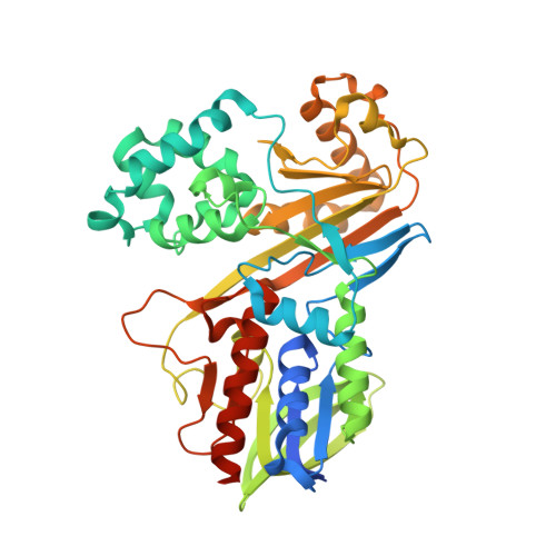
    l-Tryptophan oxidase, VioA from Chromobacterium violaceum, which has a high substrate specificity for tryptophan, is useful for quantitative assay of tryptophan. However, stability of wild type VioA is not enough for its application in clinical or industrial use. To improve the thermal stability of the enzyme, we developed a VioA (C395A) mutant, with higher stability than wild type VioA. The VioA (C395A) exhibited similar specificity and kinetic parameter for tryptophan to wild type. Conventionally, the quantity of tryptophan is determined by instrumental methods, such as high-performance liquid chromatography (HPLC) after pre-column-derivatization. Using the mutant enzyme, we succeeded in the tryptophan quantification in human plasma samples, to an accuracy of <2.9% when compared to the instrumental method, and to a precision of CV <3.2%. To analyse the improvement in storage stability and substrate specificity, we further determined the crystal structures of VioA (C395A) complexed with FAD, and with FAD and tryptophan at 1.8 Å resolution.


  • Organizational Affiliation

    Fundamental Technology Labs., Institute for Innovation, Ajinomoto Co., Inc., Kawasaki, Japan.


Macromolecules
Find similar proteins by:  (by identity cutoff)  |  3D Structure
Entity ID: 1
MoleculeChains Sequence LengthOrganismDetailsImage
Flavin-dependent L-tryptophan oxidase VioA
A, B
451Chromobacterium violaceum ATCC 12472Mutation(s): 1 
Gene Names: vioACV_3274
EC: 1.4.3.23
UniProt
Find proteins for Q9S3V1 (Chromobacterium violaceum (strain ATCC 12472 / DSM 30191 / JCM 1249 / NBRC 12614 / NCIMB 9131 / NCTC 9757))
Explore Q9S3V1 
Go to UniProtKB:  Q9S3V1
Entity Groups  
Sequence Clusters30% Identity50% Identity70% Identity90% Identity95% Identity100% Identity
UniProt GroupQ9S3V1
Sequence Annotations
Expand
  • Reference Sequence
Experimental Data & Validation

Experimental Data

  • Method: X-RAY DIFFRACTION
  • Resolution: 1.80 Å
  • R-Value Free: 0.202 
  • R-Value Work: 0.158 
  • R-Value Observed: 0.160 
  • Space Group: P 21 21 21
Unit Cell:
Length ( Å )Angle ( ˚ )
a = 87.86α = 90
b = 89.27β = 90
c = 112.7γ = 90
Software Package:
Software NamePurpose
REFMACrefinement
XSCALEdata scaling
MOLREPphasing
PDB_EXTRACTdata extraction
XDSdata reduction

Structure Validation

View Full Validation Report


Ligand Structure Quality Assessment 


Entry History 

Deposition Data

Revision History  (Full details and data files)

  • Version 1.0: 2018-12-19
    Type: Initial release
  • Version 1.1: 2024-03-27
    Changes: Data collection, Database references