5ZAB

Crystal structure of cf3-aequorin


Experimental Data Snapshot

  • Method: X-RAY DIFFRACTION
  • Resolution: 2.15 Å
  • R-Value Free: 0.230 
  • R-Value Work: 0.196 
  • R-Value Observed: 0.196 

wwPDB Validation   3D Report Full Report


Ligand Structure Quality Assessment 


This is version 1.2 of the entry. See complete history


Literature

Slow luminescence kinetics of semi-synthetic aequorin: expression, purification and structure determination of cf3-aequorin.

Inouye, S.Tomabechi, Y.Hosoya, T.Sekine, S.I.Shirouzu, M.

(2018) J Biochem 164: 247-255

  • DOI: https://doi.org/10.1093/jb/mvy049
  • Primary Citation of Related Structures:  
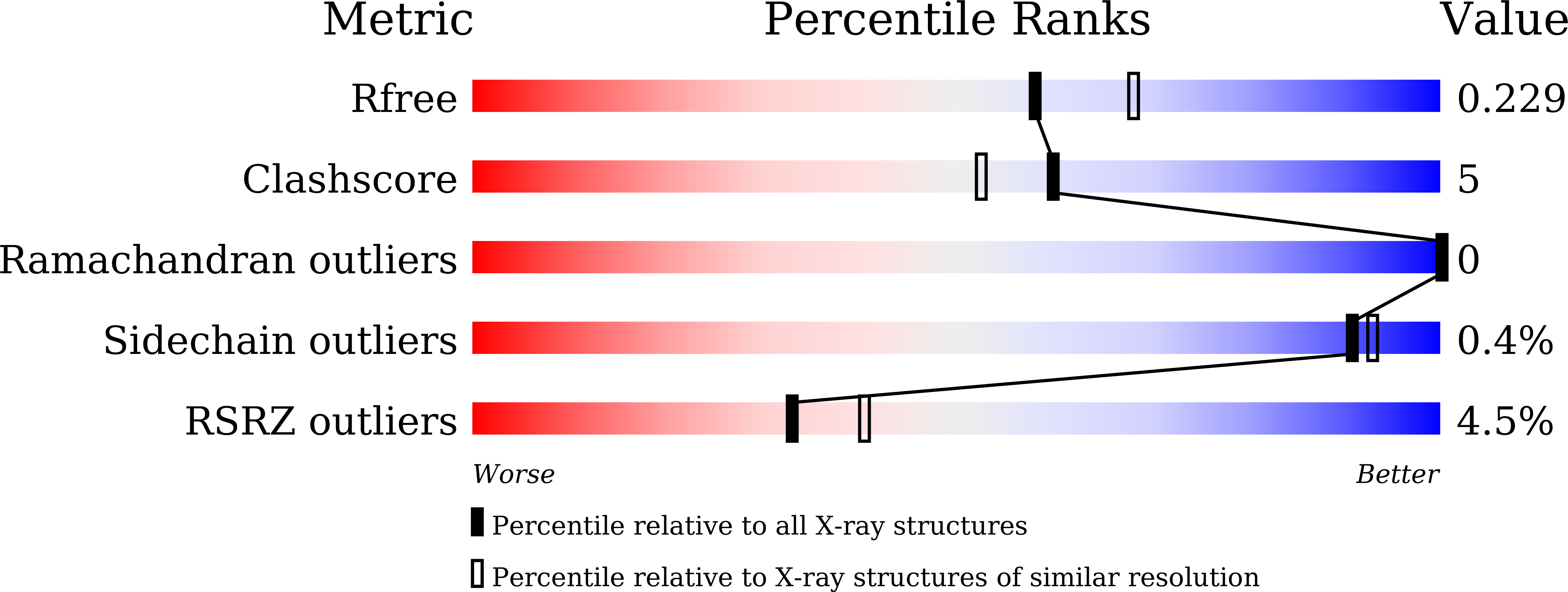
    5ZAB

  • PubMed Abstract: 

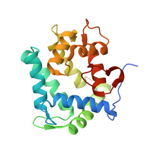
    cf3-Aequorin is one of the semi-synthetic aequorins that was produced by replacing 2-peroxycoelenterazine (CTZ-OOH) in native aequorin with a 2-peroxycoelenterazine analog, and it was prepared using the C2-modified trifluoromethyl analog of coelenterazine (cf3-CTZ) and the histidine-tagged apoaequorin expressed in Escherichia coli cells. The purified cf3-aequorin showed a slow luminescence pattern with half-decay time of maximum intensities of luminescence of 5.0 s. This is much longer than that of 0.9 s for native aequorin, and its luminescence capacity was estimated to be 72.8% of that of native aequorin. The crystal structure of cf3-aequorin was determined at 2.15 Å resolution. The light source of 2-peroxytrifluoromethylcoelenterazine (cf3-CTZ-OOH) was stabilized by the hydrogen-bonding interactions at the C2-peroxy moiety and the p-hydroxy moiety at the C6-phenyl group. In native aequorin, three water molecules contribute to stabilizing CTZ-OOH through hydrogen bonds. However, cf3-aequorin only contained one water molecule, and the trifluoromethyl moiety at the C2-benzyl group of cf3-CTZ-OOH interacted with the protein by van der Waals interactions. The slow luminescence kinetics of cf3-aequorin could be explained by slow conformational changes due to the bulkiness of the trifluoromethyl group, which might hinder the smooth cleavage of hydrogen bonds at the C2-peroxy moiety after the binding of Ca2+ to cf3-aequorin.


  • Organizational Affiliation

    Yokohama Research Center, JNC. Co., 5-1 Okawa, Kanazawa-ku, Yokohama, Japan.


Macromolecules
Find similar proteins by:  (by identity cutoff)  |  3D Structure
Entity ID: 1
MoleculeChains Sequence LengthOrganismDetailsImage
Aequorin-2
A, B, C, D, E
A, B, C, D, E, F, G, H, I, J, K, L, M, N, O, P
198Aequorea victoriaMutation(s): 0 
UniProt
Find proteins for P02592 (Aequorea victoria)
Explore P02592 
Go to UniProtKB:  P02592
Entity Groups  
Sequence Clusters30% Identity50% Identity70% Identity90% Identity95% Identity100% Identity
UniProt GroupP02592
Sequence Annotations
Expand
  • Reference Sequence
Small Molecules
Ligands 1 Unique
IDChains Name / Formula / InChI Key2D Diagram3D Interactions
9A3
Query on 9A3

Download Ideal Coordinates CCD File 
AA [auth K]
BA [auth L]
CA [auth M]
DA [auth N]
EA [auth O]
AA [auth K],
BA [auth L],
CA [auth M],
DA [auth N],
EA [auth O],
FA [auth P],
Q [auth A],
R [auth B],
S [auth C],
T [auth D],
U [auth E],
V [auth F],
W [auth G],
X [auth H],
Y [auth I],
Z [auth J]
(2S)-8-benzyl-2-hydroperoxy-6-(4-hydroxyphenyl)-2-{[4-(trifluoromethyl)phenyl]methyl}imidazo[1,2-a]pyrazin-3(2H)-one
C27 H20 F3 N3 O4
ISIFAODRJRHDGD-SANMLTNESA-N
Experimental Data & Validation

Experimental Data

  • Method: X-RAY DIFFRACTION
  • Resolution: 2.15 Å
  • R-Value Free: 0.230 
  • R-Value Work: 0.196 
  • R-Value Observed: 0.196 
  • Space Group: P 1
Unit Cell:
Length ( Å )Angle ( ˚ )
a = 91.8α = 77.62
b = 97.55β = 73.06
c = 121.84γ = 75.17
Software Package:
Software NamePurpose
XDSdata reduction
XSCALEdata scaling
PHASERphasing
PHENIXrefinement
PDB_EXTRACTdata extraction

Structure Validation

View Full Validation Report


Ligand Structure Quality Assessment 


Entry History 

Deposition Data

Revision History  (Full details and data files)

  • Version 1.0: 2018-06-06
    Type: Initial release
  • Version 1.1: 2018-08-29
    Changes: Data collection, Database references
  • Version 1.2: 2023-11-22
    Changes: Data collection, Database references, Refinement description