Structural basis for neutralization of cytotoxic abrin by monoclonal antibody D6F10.
Bansia, H., Bagaria, S., Karande, A.A., Ramakumar, S.(2019) FEBS J 286: 1003-1029
- PubMed: 30521151
- DOI: https://doi.org/10.1111/febs.14716
- Primary Citation of Related Structures:
5Z37, 5Z3I, 5Z3J - PubMed Abstract:
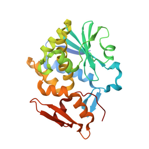
Abrin, an extremely cytotoxic Type II ribosome-inactivating protein (RIP), is a potential bio-warfare agent. Abrin A-chain (ABA) depurinates an adenosine of sarcin-ricin loop (SRL) from eukaryotic 28S rRNA, thereby arresting protein synthesis and leading to cell death. Monoclonal antibody (mAb) D6F10 is the only known antibody that neutralizes ABA's activity in cell-free systems as well as abrin's toxicity in vitro and in vivo. However, how binding of mAb D6F10 to abrin interferes with abrin's catalytic activity at ribosome is still poorly understood. To provide structural basis for mAb D6F10-mediated rescue of ribosome inactivation by abrin, we determined crystal structures of ABA with and without substrate analogs. The structures of ABA-substrate analogs and ribosome were used in an experiment-guided computational protocol, to construct the ABA-Ribosome complex. A homology model of the variable region (F v ) of mAb D6F10 was generated and docked with the apo-ABA structure to construct the ABA-D6F10 F v complex. Structural superposition of ABA common to ABA-D6F10 F v and ABA-Ribosome complexes reveals steric hindrance as the primary mechanism by which mAb D6F10 neutralizes abrin. In contrast to ABA alone, ABA bound to mAb D6F10 is unable to access the SRL on the ribosome owing to steric clashes of mAb D6F10 with the ribosome. Crystal structures of ABA also reveal a catalytic water molecule implicated in hydrolyzing N-glycosidic bond of the susceptible adenosine by RIPs. Furthermore, our strategy provides structural details of steric hindrance important for neutralization of ricin, another RIP, by mAb 6C2 and hence is of wide applicability. ENZYME: EC3.2.2.22. DATABASE: Structural data have been deposited in the Protein Data Bank (PDB) under the accession numbers 5Z37, 5Z3I, and 5Z3J.
Organizational Affiliation:
Department of Physics, Indian Institute of Science, Bengaluru, India.