5Z2L

Crystal structure of BdcA in complex with NADPH


Experimental Data Snapshot

  • Method: X-RAY DIFFRACTION
  • Resolution: 1.70 Å
  • R-Value Free: 0.179 
  • R-Value Work: 0.151 
  • R-Value Observed: 0.151 

wwPDB Validation   3D Report Full Report


Ligand Structure Quality Assessment 


This is version 1.2 of the entry. See complete history


Literature

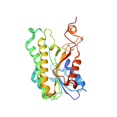
A potential substrate binding pocket of BdcA plays a critical role in NADPH recognition and biofilm dispersal

Yang, W.S.Hong, Y.Zhang, Y.Wang, D.C.Li, D.F.Hou, Y.J.

(2018) Biochem Biophys Res Commun 497: 863-868

  • DOI: https://doi.org/10.1016/j.bbrc.2018.02.143
  • Primary Citation of Related Structures:  
    5Z2L

  • PubMed Abstract: 

    Biofilm dispersal is characterized by the cell detachment from biofilms and expected to provide novel "anti-biofilm" approaches of prevention and treatment of biofilms in clinical and industrial settings. The E.coli protein BdcA has been identified as a biofilm dispersal factor and designed to be an important component in engineered applications to control biofilm formation. It belongs to short-chain dehydrogenase/reductase (SDR) family with the specific affinity to NADPH. Here, we show the structure of BdcA in complex with NADPH and confirm that NADPH binding is requisite for BdcA facilitating cell motility and increasing biofilm dispersal. Especially, we observe a potential substrate binding pocket surrounded by hydrophobic residues upon NADPH binding and present evidences that this pocket is essential for BdcA binding NADPH and exerting its biological functions. Our study provides the clues for illuminating the molecular mechanism of BdcA regulating biofilm dispersal and better utilizing BdcA to eliminate the biofilms.


  • Organizational Affiliation

    National Laboratory of Biomacromolecules, CAS Center for Excellence in Biomacromolecules, Institute of Biophysics, Chinese Academy of Sciences, Beijing 100101, China; University of Chinese Academy of Sciences, Beijing 100049, China.


Macromolecules
Find similar proteins by:  (by identity cutoff)  |  3D Structure
Entity ID: 1
MoleculeChains Sequence LengthOrganismDetailsImage
Cyclic-di-GMP-binding biofilm dispersal mediator protein245Escherichia coli K-12Mutation(s): 0 
Gene Names: bdcAyjgIb4249JW4207
UniProt
Find proteins for P39333 (Escherichia coli (strain K12))
Explore P39333 
Go to UniProtKB:  P39333
Entity Groups  
Sequence Clusters30% Identity50% Identity70% Identity90% Identity95% Identity100% Identity
UniProt GroupP39333
Sequence Annotations
Expand
  • Reference Sequence
Small Molecules
Ligands 6 Unique
IDChains Name / Formula / InChI Key2D Diagram3D Interactions
NDP (Subject of Investigation/LOI)
Query on NDP

Download Ideal Coordinates CCD File 
BA [auth K]
DA [auth I]
FA [auth F]
HA [auth G]
IA [auth L]
BA [auth K],
DA [auth I],
FA [auth F],
HA [auth G],
IA [auth L],
N [auth A],
O [auth D],
Q [auth B],
U [auth C],
X [auth E],
Y [auth J],
Z [auth H]
NADPH DIHYDRO-NICOTINAMIDE-ADENINE-DINUCLEOTIDE PHOSPHATE
C21 H30 N7 O17 P3
ACFIXJIJDZMPPO-NNYOXOHSSA-N
PG4
Query on PG4

Download Ideal Coordinates CCD File 
EA [auth F]TETRAETHYLENE GLYCOL
C8 H18 O5
UWHCKJMYHZGTIT-UHFFFAOYSA-N
PG0
Query on PG0

Download Ideal Coordinates CCD File 
M [auth A]2-(2-METHOXYETHOXY)ETHANOL
C5 H12 O3
SBASXUCJHJRPEV-UHFFFAOYSA-N
PEG
Query on PEG

Download Ideal Coordinates CCD File 
AA [auth K]DI(HYDROXYETHYL)ETHER
C4 H10 O3
MTHSVFCYNBDYFN-UHFFFAOYSA-N
GOL
Query on GOL

Download Ideal Coordinates CCD File 
R [auth C],
V [auth E]
GLYCEROL
C3 H8 O3
PEDCQBHIVMGVHV-UHFFFAOYSA-N
EDO
Query on EDO

Download Ideal Coordinates CCD File 
CA [auth I]
GA [auth G]
P [auth B]
S [auth C]
T [auth C]
CA [auth I],
GA [auth G],
P [auth B],
S [auth C],
T [auth C],
W [auth E]
1,2-ETHANEDIOL
C2 H6 O2
LYCAIKOWRPUZTN-UHFFFAOYSA-N
Binding Affinity Annotations 
IDSourceBinding Affinity
NDP Binding MOAD:  5Z2L Kd: 4.50e+4 (nM) from 1 assay(s)
Experimental Data & Validation

Experimental Data

  • Method: X-RAY DIFFRACTION
  • Resolution: 1.70 Å
  • R-Value Free: 0.179 
  • R-Value Work: 0.151 
  • R-Value Observed: 0.151 
  • Space Group: P 1
Unit Cell:
Length ( Å )Angle ( ˚ )
a = 65.833α = 99.22
b = 72.28β = 86.78
c = 172.324γ = 107.7
Software Package:
Software NamePurpose
PHENIXrefinement
XDSdata reduction
Aimlessdata scaling
PHASERphasing

Structure Validation

View Full Validation Report


Ligand Structure Quality Assessment 


Entry History & Funding Information

Deposition Data


Funding OrganizationLocationGrant Number
National Natural Science Foundation of ChinaChina31700661
National Natural Science Foundation of ChinaChina31270792

Revision History  (Full details and data files)

  • Version 1.0: 2018-03-07
    Type: Initial release
  • Version 1.1: 2018-03-21
    Changes: Database references
  • Version 1.2: 2023-11-22
    Changes: Data collection, Database references, Refinement description