5Z0Q

Crystal Structure of OvoB


Experimental Data Snapshot

  • Method: X-RAY DIFFRACTION
  • Resolution: 2.77 Å
  • R-Value Free: 0.267 
  • R-Value Work: 0.210 
  • R-Value Observed: 0.212 

wwPDB Validation   3D Report Full Report


Ligand Structure Quality Assessment 


This is version 1.1 of the entry. See complete history


Literature

In Vitro Reconstitution of the Remaining Steps in Ovothiol A Biosynthesis: C-S Lyase and Methyltransferase Reactions.

Naowarojna, N.Huang, P.Cai, Y.Song, H.Wu, L.Cheng, R.Li, Y.Wang, S.Lyu, H.Zhang, L.Zhou, J.Liu, P.

(2018) Org Lett 20: 5427-5430

  • DOI: https://doi.org/10.1021/acs.orglett.8b02332
  • Primary Citation of Related Structures:  
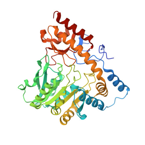
    5Z0Q

  • PubMed Abstract: 

    Ovothiols are thiolhistidine derivatives. The first step of ovothiol biosynthesis is OvoA-catalyzed oxidative coupling between histidine and cysteine. In this report, the remaining steps of ovothiol A biosynthesis were reconstituted in vitro. ETA_14770 (OvoB) was reported as a PLP-dependent sulfoxide lyase, responsible for mercaptohistidine production. OvoA was found to be a bifunctional enzyme, which mediates both oxidative C-S bond formation and methylation of mercaptohistidine to afford ovothiol A. Besides reconstituting the whole biosynthetic pathway, two unique features proposed in the literature were also examined: a potential cysteine-recycling mechanism of the C-S lyase (OvoB) and the selectivity of the π- N methyltransferase.


  • Organizational Affiliation

    Department of Chemistry , Boston University , 590 Commonwealth Avenue , Boston , Massachusetts 02215 , United States.


Macromolecules
Find similar proteins by:  (by identity cutoff)  |  3D Structure
Entity ID: 1
MoleculeChains Sequence LengthOrganismDetailsImage
Aminotransferase, class I and II389Erwinia tasmaniensis Et1/99Mutation(s): 0 
Gene Names: ETA_14770
UniProt
Find proteins for B2VJB8 (Erwinia tasmaniensis (strain DSM 17950 / CFBP 7177 / CIP 109463 / NCPPB 4357 / Et1/99))
Explore B2VJB8 
Go to UniProtKB:  B2VJB8
Entity Groups  
Sequence Clusters30% Identity50% Identity70% Identity90% Identity95% Identity100% Identity
UniProt GroupB2VJB8
Sequence Annotations
Expand
  • Reference Sequence
Small Molecules
Ligands 1 Unique
IDChains Name / Formula / InChI Key2D Diagram3D Interactions
PLP
Query on PLP

Download Ideal Coordinates CCD File 
M [auth C]
N [auth A]
O [auth B]
P [auth D]
Q [auth E]
M [auth C],
N [auth A],
O [auth B],
P [auth D],
Q [auth E],
R [auth F],
S [auth G],
T [auth H],
U [auth I],
V [auth J],
W [auth K],
X [auth L]
PYRIDOXAL-5'-PHOSPHATE
C8 H10 N O6 P
NGVDGCNFYWLIFO-UHFFFAOYSA-N
Experimental Data & Validation

Experimental Data

  • Method: X-RAY DIFFRACTION
  • Resolution: 2.77 Å
  • R-Value Free: 0.267 
  • R-Value Work: 0.210 
  • R-Value Observed: 0.212 
  • Space Group: P 1
Unit Cell:
Length ( Å )Angle ( ˚ )
a = 58.123α = 89.39
b = 133.59β = 84.58
c = 165.703γ = 86.56
Software Package:
Software NamePurpose
PHENIXrefinement
HKL-3000data reduction
HKL-3000data scaling
PHENIXphasing

Structure Validation

View Full Validation Report


Ligand Structure Quality Assessment 


Entry History 

Deposition Data

Revision History  (Full details and data files)

  • Version 1.0: 2018-11-07
    Type: Initial release
  • Version 1.1: 2023-11-22
    Changes: Data collection, Database references, Refinement description