5YPR

Crystal Structure of PSD-95 SH3-GK domain in complex with a synthesized inhibitor


Experimental Data Snapshot

  • Method: X-RAY DIFFRACTION
  • Resolution: 2.35 Å
  • R-Value Free: 0.264 
  • R-Value Work: 0.222 
  • R-Value Observed: 0.224 

wwPDB Validation   3D Report Full Report


This is version 1.2 of the entry. See complete history


Literature

Synaptic Targeting and Function of SAPAPs Mediated by Phosphorylation-Dependent Binding to PSD-95 MAGUKs.

Zhu, J.Zhou, Q.Shang, Y.Li, H.Peng, M.Ke, X.Weng, Z.Zhang, R.Huang, X.Li, S.S.C.Feng, G.Lu, Y.Zhang, M.

(2017) Cell Rep 21: 3781-3793

  • DOI: https://doi.org/10.1016/j.celrep.2017.11.107
  • Primary Citation of Related Structures:  
    5YPO, 5YPR

  • PubMed Abstract: 

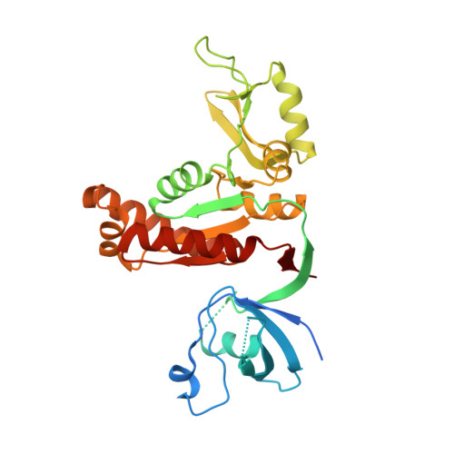
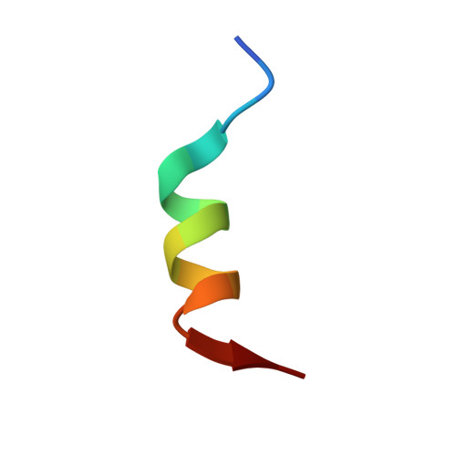
    The PSD-95/SAPAP/Shank complex functions as the major scaffold in orchestrating the formation and plasticity of the post-synaptic densities (PSDs). We previously demonstrated that the exquisitely specific SAPAP/Shank interaction is critical for Shank synaptic targeting and Shank-mediated synaptogenesis. Here, we show that the PSD-95/SAPAP interaction, SAPAP synaptic targeting, and SAPAP-mediated synaptogenesis require phosphorylation of the N-terminal repeat sequences of SAPAPs. The atomic structure of the PSD-95 guanylate kinase (GK) in complex with a phosphor-SAPAP repeat peptide, together with biochemical studies, reveals the molecular mechanism underlying the phosphorylation-dependent PSD-95/SAPAP interaction, and it also provides an explanation of a PSD-95 mutation found in patients with intellectual disabilities. Guided by the structural data, we developed potent non-phosphorylated GK inhibitory peptides capable of blocking the PSD-95/SAPAP interaction and interfering with PSD-95/SAPAP-mediated synaptic maturation and strength. These peptides are genetically encodable for investigating the functions of the PSD-95/SAPAP interaction in vivo.


  • Organizational Affiliation

    National Center for Protein Science Shanghai, CAS Center for Excellence in Molecular Cell Science, Shanghai Institute of Biochemistry and Cell Biology, Chinese Academy of Sciences, Shanghai 201203, China; University of Chinese Academy of Sciences, 333 Haike Road, Shanghai 201203, China; Division of Life Science, State Key Laboratory of Molecular Neuroscience, Hong Kong University of Science and Technology, Clear Water Bay, Kowloon, Hong Kong, China.


Macromolecules
Find similar proteins by:  (by identity cutoff)  |  3D Structure
Entity ID: 1
MoleculeChains Sequence LengthOrganismDetailsImage
Disks large homolog 4318Rattus norvegicusMutation(s): 0 
Gene Names: Dlg4Dlgh4Psd95
UniProt
Find proteins for P31016 (Rattus norvegicus)
Explore P31016 
Go to UniProtKB:  P31016
Entity Groups  
Sequence Clusters30% Identity50% Identity70% Identity90% Identity95% Identity100% Identity
UniProt GroupP31016
Sequence Annotations
Expand
  • Reference Sequence

Find similar proteins by:  Sequence   |   3D Structure  

Entity ID: 2
MoleculeChains Sequence LengthOrganismDetailsImage
Synthesized GK inhibitor16synthetic constructMutation(s): 0 
Entity Groups  
Sequence Clusters30% Identity50% Identity70% Identity90% Identity95% Identity100% Identity
Sequence Annotations
Expand
  • Reference Sequence
Experimental Data & Validation

Experimental Data

  • Method: X-RAY DIFFRACTION
  • Resolution: 2.35 Å
  • R-Value Free: 0.264 
  • R-Value Work: 0.222 
  • R-Value Observed: 0.224 
  • Space Group: C 1 2 1
Unit Cell:
Length ( Å )Angle ( ˚ )
a = 119.028α = 90
b = 45.743β = 103.22
c = 86.358γ = 90
Software Package:
Software NamePurpose
PHENIXrefinement
HKL-3000data reduction
HKL-3000data scaling
PHENIXphasing

Structure Validation

View Full Validation Report


Entry History & Funding Information

Deposition Data


Funding OrganizationLocationGrant Number
National Science Foundation (NSF, United States)China31770779, 31470733 and U1532121
Shanghai Municipal Science and Technology CommissionChina14YF1406700
RGCHong Kong16103614, 16149516 and AoE-M09-12
the Minister of Science and Technology of ChinaChina2014CB910204

Revision History  (Full details and data files)

  • Version 1.0: 2018-03-14
    Type: Initial release
  • Version 1.1: 2022-03-23
    Changes: Author supporting evidence, Database references
  • Version 1.2: 2023-11-22
    Changes: Data collection, Refinement description