


Crystal Structure of LysM domain from pteris ryukyuensis chitinase A
Kitaoku, Y., Umemoto, N., Taira, T., Fukamizo, T., Numata, T., Ohnuma, T.To be published.
Experimental Data Snapshot
wwPDB Validation 3D Report Full Report

Entity ID: 1 | |||||
|---|---|---|---|---|---|
| Molecule | Chains | Sequence Length | Organism | Details | Image |
| Chitinase A | 49 | Pteris ryukyuensis | Mutation(s): 0 Gene Names: prchiA | 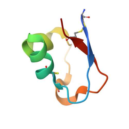 | |
UniProt | |||||
Find proteins for Q0WYK2 (Pteris ryukyuensis) Explore Q0WYK2 Go to UniProtKB: Q0WYK2 | |||||
Entity Groups | |||||
| Sequence Clusters | 30% Identity50% Identity70% Identity90% Identity95% Identity100% Identity | ||||
| UniProt Group | Q0WYK2 | ||||
Sequence AnnotationsExpand | |||||
| |||||
| Ligands 2 Unique | |||||
|---|---|---|---|---|---|
| ID | Chains | Name / Formula / InChI Key | 2D Diagram | 3D Interactions | |
| ZN Query on ZN | E [auth A], F [auth A], H [auth A] | ZINC ION Zn PTFCDOFLOPIGGS-UHFFFAOYSA-N |  | ||
| EDO Query on EDO | G [auth A], I [auth D] | 1,2-ETHANEDIOL C2 H6 O2 LYCAIKOWRPUZTN-UHFFFAOYSA-N |  | ||
| Length ( Å ) | Angle ( ˚ ) |
|---|---|
| a = 38.839 | α = 90 |
| b = 50.125 | β = 90 |
| c = 92.362 | γ = 90 |
| Software Name | Purpose |
|---|---|
| REFMAC | refinement |
| HKL-2000 | data scaling |
| Coot | model building |
| PHASER | phasing |
| DENZO | data reduction |
| Funding Organization | Location | Grant Number |
|---|---|---|
| MEXT (Ministry of Education, Culture, Sports, Science, and Technology) | Japan | S1101035 |
RCSB PDB (citation) is hosted by
RCSB PDB is a member of the

