5YJ8

Identification of a small molecule inhibitor for the Tudor domain of TDRD3


Experimental Data Snapshot

  • Method: X-RAY DIFFRACTION
  • Resolution: 1.76 Å
  • R-Value Free: 0.226 
  • R-Value Work: 0.187 
  • R-Value Observed: 0.191 

wwPDB Validation   3D Report Full Report


Ligand Structure Quality Assessment 


This is version 1.3 of the entry. See complete history


Literature

Structural plasticity of the TDRD3 Tudor domain probed by a fragment screening hit.

Liu, J.Zhang, S.Liu, M.Liu, Y.Nshogoza, G.Gao, J.Ma, R.Yang, Y.Wu, J.Zhang, J.Li, F.Ruan, K.

(2018) FEBS J 285: 2091-2103

  • DOI: https://doi.org/10.1111/febs.14469
  • Primary Citation of Related Structures:  
    5YJ8

  • PubMed Abstract: 

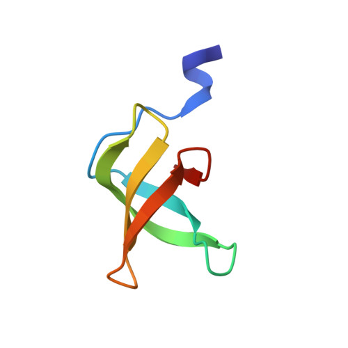
    As a reader of di-methylated arginine on various proteins, such as histone, RNA polymerase II, PIWI and Fragile X mental retardation protein, the Tudor domain of Tudor domain-containing protein 3 (TDRD3) mediates transcriptional activation in nucleus and formation of stress granules in the cytoplasm. Despite the TDRD3 implication in cancer cell proliferation and invasion, warheads to block the di-methylated arginine recognition pocket of the TDRD3 Tudor domain have not yet been uncovered. Here we identified 14 small molecule hits against the TDRD3 Tudor domain through NMR fragment-based screening. These hits were further cross-validated by using competitive fluorescence polarization and isothermal titration calorimetry experiments. The crystal structure of the TDRD3 Tudor domain in complex with hit 1 reveals a distinct binding mode from the nature substrate. Hit 1 protrudes into the aromatic cage of the TDRD3 Tudor domain, where the aromatic residues are tilted to accommodate a sandwich-like π-π interaction. The side chain of the conserved residue N596 swings away 3.1 Å to form a direct hydrogen bond with hit 1. Moreover, this compound shows a decreased affinity against the single Tudor domain of survival motor neuron protein, but no detectable binding to neither the tandem Tudor domain of TP53-binding protein 1 nor the extended Tudor domain of staphylococcal nuclease domain-containing protein 1. Our work depicts the structural plasticity of the TDRD3 Tudor domain and paves the way for the subsequent structure-guided discovery of selective inhibitors targeting Tudor domains.


  • Organizational Affiliation

    Hefei National Laboratory for Physical Sciences at the Microscale, School of Life Sciences, University of Science and Technology of China, Hefei, China.


Macromolecules
Find similar proteins by:  (by identity cutoff)  |  3D Structure
Entity ID: 1
MoleculeChains Sequence LengthOrganismDetailsImage
Tudor domain-containing protein 360Homo sapiensMutation(s): 0 
Gene Names: TDRD3
UniProt & NIH Common Fund Data Resources
Find proteins for Q9H7E2 (Homo sapiens)
Explore Q9H7E2 
Go to UniProtKB:  Q9H7E2
PHAROS:  Q9H7E2
GTEx:  ENSG00000083544 
Entity Groups  
Sequence Clusters30% Identity50% Identity70% Identity90% Identity95% Identity100% Identity
UniProt GroupQ9H7E2
Sequence Annotations
Expand
  • Reference Sequence
Small Molecules
Ligands 2 Unique
IDChains Name / Formula / InChI Key2D Diagram3D Interactions
8W9
Query on 8W9

Download Ideal Coordinates CCD File 
B [auth A]5-(aminomethyl)-1,3-dimethyl-benzimidazol-2-one
C10 H13 N3 O
GDVGETHSXBLPHM-UHFFFAOYSA-N
SO4
Query on SO4

Download Ideal Coordinates CCD File 
C [auth A]SULFATE ION
O4 S
QAOWNCQODCNURD-UHFFFAOYSA-L
Binding Affinity Annotations 
IDSourceBinding Affinity
8W9 Binding MOAD:  5YJ8 Kd: 1.04e+5 (nM) from 1 assay(s)
Experimental Data & Validation

Experimental Data

  • Method: X-RAY DIFFRACTION
  • Resolution: 1.76 Å
  • R-Value Free: 0.226 
  • R-Value Work: 0.187 
  • R-Value Observed: 0.191 
  • Space Group: P 32 2 1
Unit Cell:
Length ( Å )Angle ( ˚ )
a = 50.974α = 90
b = 50.974β = 90
c = 42.087γ = 120
Software Package:
Software NamePurpose
PHENIXrefinement
SCALEPACKdata scaling
PDB_EXTRACTdata extraction
HKL-2000data reduction
MOLREPphasing

Structure Validation

View Full Validation Report


Ligand Structure Quality Assessment 


Entry History 

Deposition Data

Revision History  (Full details and data files)

  • Version 1.0: 2018-05-02
    Type: Initial release
  • Version 1.1: 2018-05-16
    Changes: Data collection, Database references
  • Version 1.2: 2018-06-20
    Changes: Data collection, Database references
  • Version 1.3: 2023-11-22
    Changes: Data collection, Database references, Refinement description