5Y41

Crystal Structure of LIGAND-BOUND NURR1-LBD


Experimental Data Snapshot

  • Method: X-RAY DIFFRACTION
  • Resolution: 2.05 Å
  • R-Value Free: 0.253 
  • R-Value Work: 0.206 
  • R-Value Observed: 0.207 

wwPDB Validation   3D Report Full Report


Ligand Structure Quality Assessment 


This is version 1.2 of the entry. See complete history


Literature

PGE1 and PGA1 bind to Nurr1 and activate its transcriptional function.

Rajan, S.Jang, Y.Kim, C.H.Kim, W.Toh, H.T.Jeon, J.Song, B.Serra, A.Lescar, J.Yoo, J.Y.Beldar, S.Ye, H.Kang, C.Liu, X.W.Feitosa, M.Kim, Y.Hwang, D.Goh, G.Lim, K.L.Park, H.M.Lee, C.H.Oh, S.F.Petsko, G.A.Yoon, H.S.Kim, K.S.

(2020) Nat Chem Biol 

  • DOI: https://doi.org/10.1038/s41589-020-0553-6
  • Primary Citation of Related Structures:  
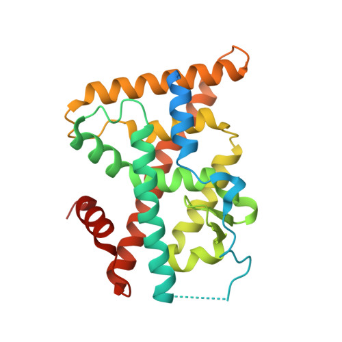
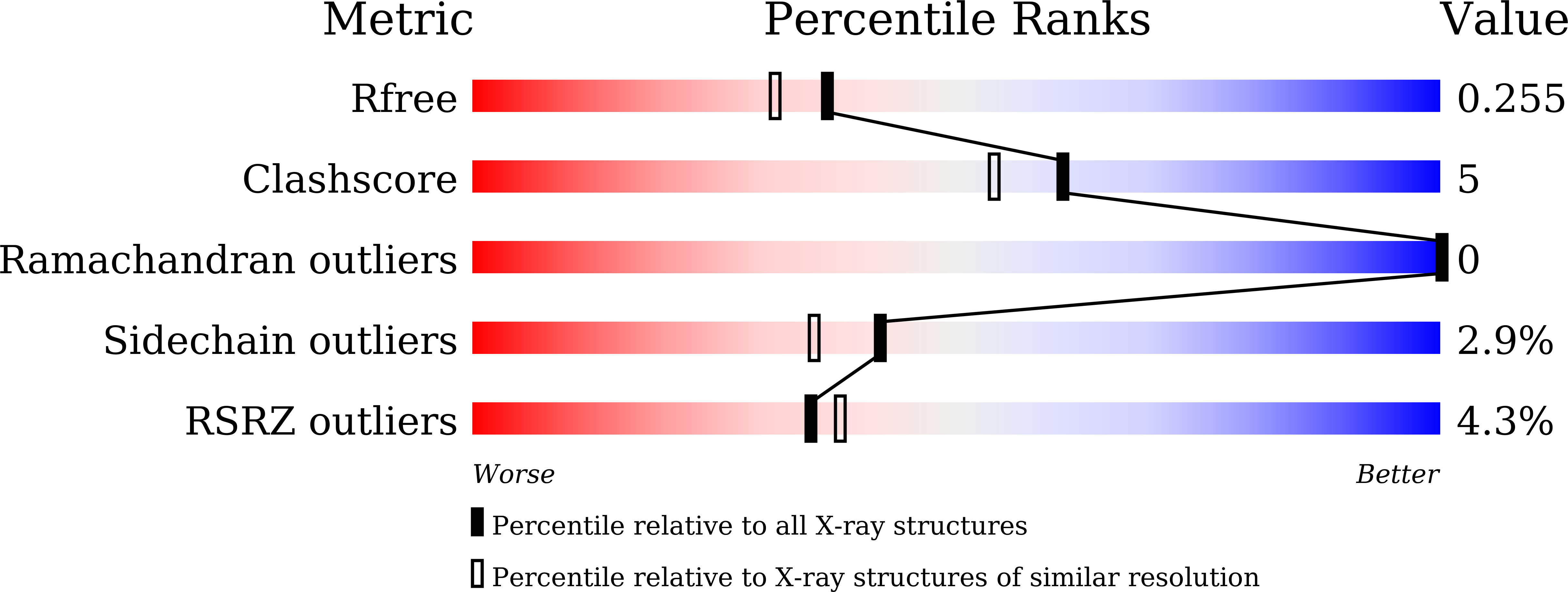
    5Y41

  • PubMed Abstract: 

    The orphan nuclear receptor Nurr1 is critical for the development, maintenance and protection of midbrain dopaminergic (mDA) neurons. Here we show that prostaglandin E1 (PGE1) and its dehydrated metabolite, PGA1, directly interact with the ligand-binding domain (LBD) of Nurr1 and stimulate its transcriptional function. We also report the crystallographic structure of Nurr1-LBD bound to PGA1 at 2.05 Å resolution. PGA1 couples covalently to Nurr1-LBD by forming a Michael adduct with Cys566, and induces notable conformational changes, including a 21° shift of the activation function-2 helix (H12) away from the protein core. Furthermore, PGE1/PGA1 exhibit neuroprotective effects in a Nurr1-dependent manner, prominently enhance expression of Nurr1 target genes in mDA neurons and improve motor deficits in 1-methyl-4-phenyl-1,2,3,6-tetrahydropyridine-lesioned mouse models of Parkinson's disease. Based on these results, we propose that PGE1/PGA1 represent native ligands of Nurr1 and can exert neuroprotective effects on mDA neurons, via activation of Nurr1's transcriptional function.


  • Organizational Affiliation

    School of Biological Sciences, Nanyang Technological University, Singapore, Singapore.


Macromolecules
Find similar proteins by:  (by identity cutoff)  |  3D Structure
Entity ID: 1
MoleculeChains Sequence LengthOrganismDetailsImage
Nuclear receptor subfamily 4 group A member 2
A, B
271Homo sapiensMutation(s): 0 
Gene Names: NR4A2NOTNURR1TINUR
UniProt & NIH Common Fund Data Resources
Find proteins for P43354 (Homo sapiens)
Explore P43354 
Go to UniProtKB:  P43354
PHAROS:  P43354
GTEx:  ENSG00000153234 
Entity Groups  
Sequence Clusters30% Identity50% Identity70% Identity90% Identity95% Identity100% Identity
UniProt GroupP43354
Sequence Annotations
Expand
  • Reference Sequence
Small Molecules
Ligands 5 Unique
IDChains Name / Formula / InChI Key2D Diagram3D Interactions
RPG
Query on RPG

Download Ideal Coordinates CCD File 
C [auth A],
H [auth B]
(13E,15S)-15-hydroxy-9-oxoprosta-10,13-dien-1-oic acid
C20 H32 O4
BGKHCLZFGPIKKU-LDDQNKHRSA-N
PGE
Query on PGE

Download Ideal Coordinates CCD File 
D [auth A]TRIETHYLENE GLYCOL
C6 H14 O4
ZIBGPFATKBEMQZ-UHFFFAOYSA-N
PEG
Query on PEG

Download Ideal Coordinates CCD File 
E [auth A]DI(HYDROXYETHYL)ETHER
C4 H10 O3
MTHSVFCYNBDYFN-UHFFFAOYSA-N
CL
Query on CL

Download Ideal Coordinates CCD File 
F [auth A],
I [auth B]
CHLORIDE ION
Cl
VEXZGXHMUGYJMC-UHFFFAOYSA-M
MG
Query on MG

Download Ideal Coordinates CCD File 
G [auth A]MAGNESIUM ION
Mg
JLVVSXFLKOJNIY-UHFFFAOYSA-N
Experimental Data & Validation

Experimental Data

  • Method: X-RAY DIFFRACTION
  • Resolution: 2.05 Å
  • R-Value Free: 0.253 
  • R-Value Work: 0.206 
  • R-Value Observed: 0.207 
  • Space Group: P 21 21 2
Unit Cell:
Length ( Å )Angle ( ˚ )
a = 88.52α = 90
b = 131.46β = 90
c = 47.13γ = 90
Software Package:
Software NamePurpose
REFMACrefinement
iMOSFLMdata reduction
SCALAdata scaling
PHASERphasing

Structure Validation

View Full Validation Report


Ligand Structure Quality Assessment 


Entry History 

Deposition Data

Revision History  (Full details and data files)

  • Version 1.0: 2018-12-26
    Type: Initial release
  • Version 1.1: 2020-06-10
    Changes: Database references
  • Version 1.2: 2023-11-22
    Changes: Data collection, Database references, Derived calculations, Refinement description