5XQG

Crystal structure of a PL 26 exo-rhamnogalacturonan lyase from Penicillium chrysogenum complexed with unsaturated galacturonosyl rhamnose


Experimental Data Snapshot

  • Method: X-RAY DIFFRACTION
  • Resolution: 2.74 Å
  • R-Value Free: 0.247 
  • R-Value Work: 0.188 
  • R-Value Observed: 0.191 

wwPDB Validation   3D Report Full Report


This is version 2.1 of the entry. See complete history


Literature

Crystal structure of exo-rhamnogalacturonan lyase from Penicillium chrysogenum as a member of polysaccharide lyase family 26

Kunishige, Y.Iwai, M.Nakazawa, M.Ueda, M.Tada, T.Nishimura, S.Sakamoto, T.

(2018) FEBS Lett 592: 1378-1388

  • DOI: https://doi.org/10.1002/1873-3468.13034
  • Primary Citation of Related Structures:  
    5XQ3, 5XQG, 5XQJ, 5XQO

  • PubMed Abstract: 

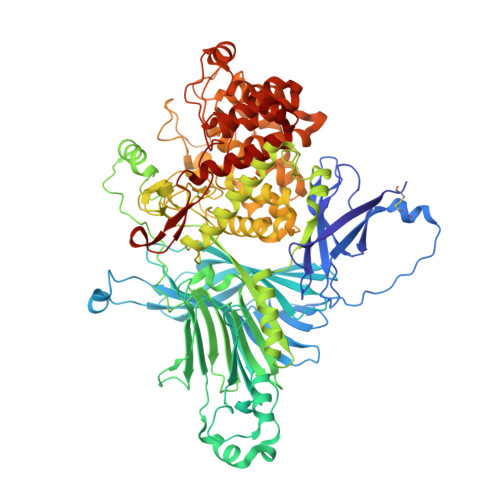
    Exo-rhamnogalacturonan lyase from Penicillium chrysogenum 31B (PcRGLX) was recently classified as a member of polysaccharide lyase (PL) family 26 along with hypothetical proteins derived from various organisms. In this study, we determined the crystal structure of PcRGLX as the first structure of a member of this family. Based on the substrate-binding orientation and substrate specificity, PcRGLX is an exo-type PL that cleaves rhamnogalacturonan from the reducing end. Analysis of PcRGLX-complex structures with reaction products indicate that the active site possesses an L-shaped cleft that can accommodate galactosyl side chains, suggesting side-chain-bypassing activity in PcRGLX. Furthermore, we determined the residues critical for catalysis by analyzing the enzyme activities of inactive variants.


  • Organizational Affiliation

    Division of Applied Life Sciences, Graduate School of Life and Environmental Sciences, Osaka Prefecture University, Sakai, Japan.


Macromolecules
Find similar proteins by:  (by identity cutoff)  |  3D Structure
Entity ID: 1
MoleculeChains Sequence LengthOrganismDetailsImage
Pcrglx protein
A, B, C, D, E
A, B, C, D, E, F, G, H
906Penicillium chrysogenumMutation(s): 0 
Gene Names: PcrglxEN45_041150
UniProt
Find proteins for B6H7Q7 (Penicillium rubens (strain ATCC 28089 / DSM 1075 / NRRL 1951 / Wisconsin 54-1255))
Explore B6H7Q7 
Go to UniProtKB:  B6H7Q7
Entity Groups  
Sequence Clusters30% Identity50% Identity70% Identity90% Identity95% Identity100% Identity
UniProt GroupB6H7Q7
Sequence Annotations
Expand
  • Reference Sequence
Oligosaccharides

Help

Entity ID: 2
MoleculeChains Length2D Diagram Glycosylation3D Interactions
2,6-anhydro-3-deoxy-L-threo-hex-2-enonic acid-(1-2)-alpha-L-rhamnopyranose
I, J, K, L, M
I, J, K, L, M, N, O, P
2N/A
Glycosylation Resources
GlyTouCan:  G94370ME
GlyCosmos:  G94370ME
Experimental Data & Validation

Experimental Data

  • Method: X-RAY DIFFRACTION
  • Resolution: 2.74 Å
  • R-Value Free: 0.247 
  • R-Value Work: 0.188 
  • R-Value Observed: 0.191 
  • Space Group: P 21 21 21
Unit Cell:
Length ( Å )Angle ( ˚ )
a = 167.878α = 90
b = 171.935β = 90
c = 342.38γ = 90
Software Package:
Software NamePurpose
REFMACrefinement
HKL-2000data collection
HKL-2000data reduction
HKL-2000data scaling
MOLREPphasing

Structure Validation

View Full Validation Report


Entry History 

Deposition Data

Revision History  (Full details and data files)

  • Version 1.0: 2018-03-21
    Type: Initial release
  • Version 1.1: 2018-04-11
    Changes: Data collection, Database references
  • Version 1.2: 2018-05-16
    Changes: Data collection, Database references, Source and taxonomy
  • Version 2.0: 2020-07-29
    Type: Remediation
    Reason: Carbohydrate remediation
    Changes: Advisory, Atomic model, Data collection, Derived calculations, Structure summary
  • Version 2.1: 2023-11-22
    Changes: Data collection, Database references, Derived calculations, Refinement description, Structure summary