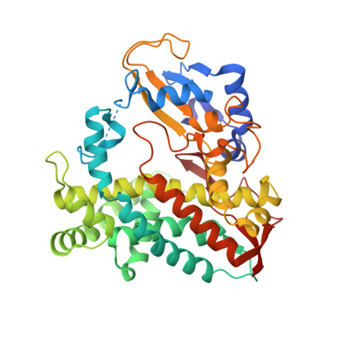
Crystal Structure and Functional Characterization of a Cytochrome P450 (BaCYP106A2) fromBacillussp. PAMC 23377.
Kim, K.H., Lee, C.W., Dangi, B., Park, S.H., Park, H., Oh, T.J., Lee, J.H.(2017) J Microbiol Biotechnol 27: 1472-1482
- PubMed: 28633515
- DOI: https://doi.org/10.4014/jmb.1706.06013
- Primary Citation of Related Structures:
5XNT - PubMed Abstract:
Bacterial cytochrome P450 (CYP) steroid hydroxylases are effectively useful in the pharmaceutical industry for introducing hydroxyl groups to a wide range of steroids. We found a putative CYP steroid hydroxylase ( Ba CYP106A2) from the bacterium Bacillus sp. PAMC 23377 isolated from Kara Sea of the Arctic Ocean, showing 94% sequence similarity with Bm CYP106A2 ( Bacillus megaterium ATCC 13368). In this study, soluble Ba CYP106A2 was overexpressed to evaluate its substrate-binding activity. The substrate affinity ( K d value) to 4-androstenedione was 387 ± 37 µM. Moreover, the crystal structure of Ba CYP106A2 was determined at 2.7 Å resolution. Structural analysis suggested that the α8-α9 loop region of Ba CYP106A2 is intrinsically mobile and might be important for initial ligand binding. The hydroxyl activity of Ba CYP106A2 was identified using in vitro enzyme assays. Its activity was confirmed with two kinds of steroid substrates, 4-androstenedione and nandrolone, using chromatography and mass spectrometry methods. The main products were monohydroxylated compounds with high conversion yields. This is the second study on the structure of CYP106A steroid hydroxylases, and should contribute new insight into the interactions of bacterial CYP106A with steroid substrates, providing baseline data for studying the CYP106A steroid hydroxylase from the structural and enzymatic perspectives.
Organizational Affiliation:
Department of Life Science and Biochemical Engineering, Sunmoon University, Asan 31460, Republic of Korea.