5XEB

Structure of the envelope glycoprotein of Dhori virus


Experimental Data Snapshot

  • Method: X-RAY DIFFRACTION
  • Resolution: 2.50 Å
  • R-Value Free: 0.257 
  • R-Value Work: 0.231 
  • R-Value Observed: 0.231 

wwPDB Validation   3D Report Full Report


Ligand Structure Quality Assessment 


This is version 1.3 of the entry. See complete history


Literature

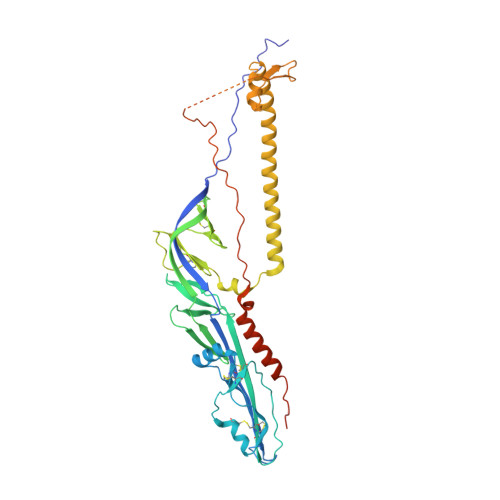
Structures of human-infectingThogotovirusfusogens support a common ancestor with insect baculovirus

Peng, R.Zhang, S.Cui, Y.Shi, Y.Gao, G.F.Qi, J.

(2017) Proc Natl Acad Sci U S A 114: E8905-E8912

  • DOI: https://doi.org/10.1073/pnas.1706125114
  • Primary Citation of Related Structures:  
    5XEA, 5XEB

  • PubMed Abstract: 

    Thogotoviruses are emerging tick-borne zoonotic orthomyxoviruses infecting both humans and domestic animals with severe clinical consequences. These viruses utilize a single-envelope glycoprotein (Gp) to facilitate their entry into host cells. Here, we present the Gp structures of Thogoto and Dhori viruses, both of which are members of the Thogotovirus genus in the family Orthomyxoviridae These structures, determined in the postfusion conformation, identified them as class III viral fusion proteins. It is intriguing that the Gp structures are similar to the envelope protein of baculovirus, although sharing a low sequence identity of ∼28%. Detailed structural and phylogenic analyses demonstrated that these Gps originated from a common ancestor. Among the structures, domain I is the most conserved region, particularly the fusion loops. Domain II showed the highest variability among different viruses, which might be related to their distinct host tropism. These findings increase our understanding of the divergent evolution processes of various orthomyxoviruses and indicate potential targets for developing antiviral therapeutics by intercepting virus entry.


  • Organizational Affiliation

    Chinese Academy of Sciences Key Laboratory of Pathogenic Microbiology and Immunology, Institute of Microbiology, Chinese Academy of Sciences, Beijing 100101, China.


Macromolecules
Find similar proteins by:  (by identity cutoff)  |  3D Structure
Entity ID: 1
MoleculeChains Sequence LengthOrganismDetailsImage
Envelope glycoprotein
A, B, C
474Dhori virus (strain Indian/1313/61)Mutation(s): 0 
Gene Names: P4
UniProt
Find proteins for P27427 (Dhori virus (strain Indian/1313/61))
Explore P27427 
Go to UniProtKB:  P27427
Entity Groups  
Sequence Clusters30% Identity50% Identity70% Identity90% Identity95% Identity100% Identity
UniProt GroupP27427
Sequence Annotations
Expand
  • Reference Sequence
Experimental Data & Validation

Experimental Data

  • Method: X-RAY DIFFRACTION
  • Resolution: 2.50 Å
  • R-Value Free: 0.257 
  • R-Value Work: 0.231 
  • R-Value Observed: 0.231 
  • Space Group: P 3
Unit Cell:
Length ( Å )Angle ( ˚ )
a = 106.729α = 90
b = 106.729β = 90
c = 134.44γ = 120
Software Package:
Software NamePurpose
PHENIXrefinement
HKL-2000data reduction
SCALEPACKdata scaling
PHASERphasing

Structure Validation

View Full Validation Report


Ligand Structure Quality Assessment 


Entry History & Funding Information

Deposition Data


Funding OrganizationLocationGrant Number
the National Natural Science Foundation of ChinaChina81641001

Revision History  (Full details and data files)

  • Version 1.0: 2017-10-04
    Type: Initial release
  • Version 1.1: 2017-11-15
    Changes: Database references
  • Version 1.2: 2018-06-13
    Changes: Data collection, Database references, Structure summary
  • Version 1.3: 2020-07-29
    Type: Remediation
    Reason: Carbohydrate remediation
    Changes: Data collection, Derived calculations, Structure summary