5WKA

Crystal structure of a GH1 beta-glucosidase retrieved from microbial metagenome of Poraque Amazon lake


Experimental Data Snapshot

  • Method: X-RAY DIFFRACTION
  • Resolution: 2.75 Å
  • R-Value Free: 0.270 
  • R-Value Work: 0.232 
  • R-Value Observed: 0.234 

wwPDB Validation   3D Report Full Report


This is version 1.4 of the entry. See complete history


Literature

A novel beta-glucosidase isolated from the microbial metagenome of Lake Poraque (Amazon, Brazil).

Toyama, D.de Morais, M.A.B.Ramos, F.C.Zanphorlin, L.M.Tonoli, C.C.C.Balula, A.F.de Miranda, F.P.Almeida, V.M.Marana, S.R.Ruller, R.Murakami, M.T.Henrique-Silva, F.

(2018) Biochim Biophys Acta 1866: 569-579

  • DOI: https://doi.org/10.1016/j.bbapap.2018.02.001
  • Primary Citation of Related Structures:  
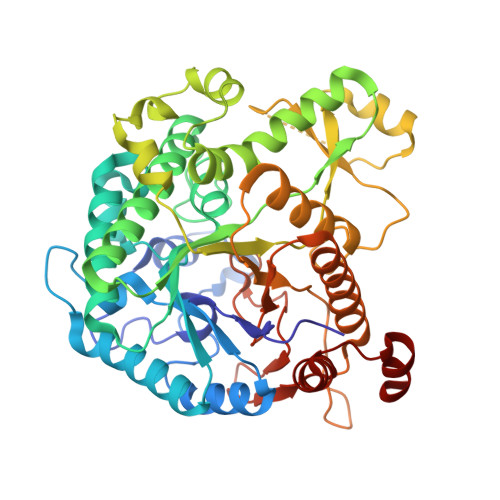
    5WKA

  • PubMed Abstract: 

    The Amazon region holds most of the biological richness of Brazil. Despite their ecological and biotechnological importance, studies related to microorganisms from this region are limited. Metagenomics leads to exciting discoveries, mainly regarding non-cultivable microorganisms. Herein, we report the discovery of a novel β-glucosidase (glycoside hydrolase family 1) gene from a metagenome from Lake Poraquê in the Amazon region. The gene encodes a protein of 52.9 kDa, named AmBgl-LP, which was recombinantly expressed in Escherichia coli and biochemically and structurally characterized. Although AmBgl-LP hydrolyzed the synthetic substrate p-nitrophenyl-β-d-glucopyranoside (pNPβG) and the natural substrate cellobiose, it showed higher specificity for pNPβG (k cat /K m  = 6 s -1 ·mM -1 ) than cellobiose (k cat /K m  = 0.6 s -1 ·mM -1 ). AmBgl-LP showed maximum activity at 40 °C and pH 6.0 when pNPβG was used as the substrate. Glucose is a competitive inhibitor of AmBgl-LP, presenting a K i of 14 mM. X-ray crystallography and Small Angle X-ray Scattering were used to determine the AmBgl-LP three-dimensional structure and its oligomeric state. Interestingly, despite sharing similar active site architecture with other structurally characterized GH1 family members which are monomeric, AmBgl-LP forms stable dimers in solution. The identification of new GH1 members by metagenomics might extend our understanding of the molecular mechanisms and diversity of these enzymes, besides enabling us to survey their industrial applications.


  • Organizational Affiliation

    Laboratory of Molecular Biology, Department of Genetics and Evolution, Federal University of São Carlos, SP, Brazil.


Macromolecules
Find similar proteins by:  (by identity cutoff)  |  3D Structure
Entity ID: 1
MoleculeChains Sequence LengthOrganismDetailsImage
Beta-glucosidase
A, B, C, D
473metagenomeMutation(s): 0 
Entity Groups  
Sequence Clusters30% Identity50% Identity70% Identity90% Identity95% Identity100% Identity
Sequence Annotations
Expand
  • Reference Sequence
Small Molecules
Ligands 2 Unique
IDChains Name / Formula / InChI Key2D Diagram3D Interactions
PEG
Query on PEG

Download Ideal Coordinates CCD File 
F [auth A],
H [auth B],
K [auth C],
N [auth D]
DI(HYDROXYETHYL)ETHER
C4 H10 O3
MTHSVFCYNBDYFN-UHFFFAOYSA-N
GOL
Query on GOL

Download Ideal Coordinates CCD File 
E [auth A]
G [auth B]
I [auth C]
J [auth C]
L [auth D]
E [auth A],
G [auth B],
I [auth C],
J [auth C],
L [auth D],
M [auth D]
GLYCEROL
C3 H8 O3
PEDCQBHIVMGVHV-UHFFFAOYSA-N
Experimental Data & Validation

Experimental Data

  • Method: X-RAY DIFFRACTION
  • Resolution: 2.75 Å
  • R-Value Free: 0.270 
  • R-Value Work: 0.232 
  • R-Value Observed: 0.234 
  • Space Group: C 1 2 1
Unit Cell:
Length ( Å )Angle ( ˚ )
a = 165.471α = 90
b = 56.652β = 96.66
c = 209.546γ = 90
Software Package:
Software NamePurpose
XSCALEdata scaling
PHENIXrefinement
PDB_EXTRACTdata extraction
XDSdata reduction
MOLREPphasing

Structure Validation

View Full Validation Report


Entry History & Funding Information

Deposition Data


Funding OrganizationLocationGrant Number
Sao Paulo Research Foundation (FAPESP)Brazil15/26982-0

Revision History  (Full details and data files)

  • Version 1.0: 2018-03-07
    Type: Initial release
  • Version 1.1: 2018-03-28
    Changes: Data collection, Database references
  • Version 1.2: 2019-04-17
    Changes: Author supporting evidence, Data collection
  • Version 1.3: 2020-01-01
    Changes: Author supporting evidence
  • Version 1.4: 2023-10-04
    Changes: Data collection, Database references, Refinement description