5W53

Crystal structure of the erythrocyte-binding domain from Plasmodium vivax reticulocyte-binding protein 2b (PvRBP2b)


Experimental Data Snapshot

  • Method: X-RAY DIFFRACTION
  • Resolution: 1.71 Å
  • R-Value Free: 0.188 
  • R-Value Work: 0.168 
  • R-Value Observed: 0.168 

wwPDB Validation   3D Report Full Report


This is version 1.2 of the entry. See complete history


Literature

Transferrin receptor 1 is a reticulocyte-specific receptor for Plasmodium vivax.

Gruszczyk, J.Kanjee, U.Chan, L.J.Menant, S.Malleret, B.Lim, N.T.Y.Schmidt, C.Q.Mok, Y.F.Lin, K.M.Pearson, R.D.Rangel, G.Smith, B.J.Call, M.J.Weekes, M.P.Griffin, M.D.W.Murphy, J.M.Abraham, J.Sriprawat, K.Menezes, M.J.Ferreira, M.U.Russell, B.Renia, L.Duraisingh, M.T.Tham, W.H.

(2018) Science 359: 48-55

  • DOI: https://doi.org/10.1126/science.aan1078
  • Primary Citation of Related Structures:  
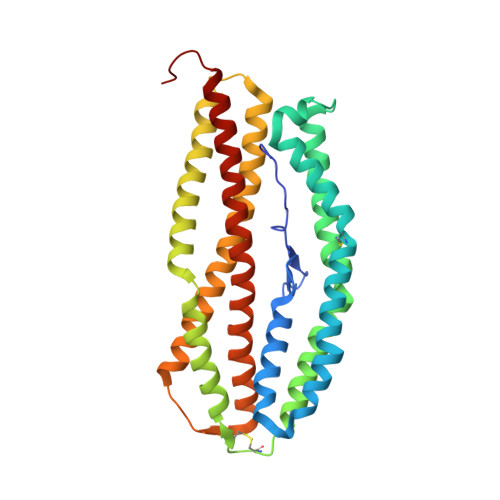
    5W53

  • PubMed Abstract: 

    Plasmodium vivax shows a strict host tropism for reticulocytes. We identified transferrin receptor 1 (TfR1) as the receptor for P. vivax reticulocyte-binding protein 2b (PvRBP2b). We determined the structure of the N-terminal domain of PvRBP2b involved in red blood cell binding, elucidating the molecular basis for TfR1 recognition. We validated TfR1 as the biological target of PvRBP2b engagement by means of TfR1 expression knockdown analysis. TfR1 mutant cells deficient in PvRBP2b binding were refractory to invasion of P. vivax but not to invasion of P. falciparum Using Brazilian and Thai clinical isolates, we show that PvRBP2b monoclonal antibodies that inhibit reticulocyte binding also block P. vivax entry into reticulocytes. These data show that TfR1-PvRBP2b invasion pathway is critical for the recognition of reticulocytes during P. vivax invasion.


  • Organizational Affiliation

    The Walter and Eliza Hall Institute of Medical Research, Parkville, Victoria 3052, Australia.


Macromolecules
Find similar proteins by:  (by identity cutoff)  |  3D Structure
Entity ID: 1
MoleculeChains Sequence LengthOrganismDetailsImage
Reticulocyte binding protein 2, putative
A, B
307Plasmodium vivax Sal-1Mutation(s): 0 
Gene Names: PVX_094255
UniProt
Find proteins for A5K736 (Plasmodium vivax (strain Salvador I))
Explore A5K736 
Go to UniProtKB:  A5K736
Entity Groups  
Sequence Clusters30% Identity50% Identity70% Identity90% Identity95% Identity100% Identity
UniProt GroupA5K736
Sequence Annotations
Expand
  • Reference Sequence
Small Molecules
Ligands 2 Unique
IDChains Name / Formula / InChI Key2D Diagram3D Interactions
SCN
Query on SCN

Download Ideal Coordinates CCD File 
C [auth A],
D [auth A],
E [auth A],
K [auth B],
L [auth B]
THIOCYANATE ION
C N S
ZMZDMBWJUHKJPS-UHFFFAOYSA-M
K
Query on K

Download Ideal Coordinates CCD File 
F [auth A]
G [auth A]
H [auth A]
I [auth A]
J [auth B]
F [auth A],
G [auth A],
H [auth A],
I [auth A],
J [auth B],
M [auth B],
N [auth B],
O [auth B]
POTASSIUM ION
K
NPYPAHLBTDXSSS-UHFFFAOYSA-N
Experimental Data & Validation

Experimental Data

  • Method: X-RAY DIFFRACTION
  • Resolution: 1.71 Å
  • R-Value Free: 0.188 
  • R-Value Work: 0.168 
  • R-Value Observed: 0.168 
  • Space Group: P 1 21 1
Unit Cell:
Length ( Å )Angle ( ˚ )
a = 59.5α = 90
b = 124.2β = 97.75
c = 65.31γ = 90
Software Package:
Software NamePurpose
iMOSFLMdata reduction
Aimlessdata scaling
PHASERphasing
PHENIXrefinement

Structure Validation

View Full Validation Report


Entry History 

Deposition Data

Revision History  (Full details and data files)

  • Version 1.0: 2017-11-29
    Type: Initial release
  • Version 1.1: 2018-01-17
    Changes: Database references
  • Version 1.2: 2023-10-04
    Changes: Data collection, Database references, Derived calculations, Refinement description