5V90

Crystal structure of ERp29 D-domain in complex with the P-domain of calreticulin


Experimental Data Snapshot

  • Method: X-RAY DIFFRACTION
  • Resolution: 3.25 Å
  • R-Value Free: 0.262 
  • R-Value Work: 0.240 
  • R-Value Observed: 0.241 

wwPDB Validation   3D Report Full Report


This is version 1.4 of the entry. See complete history


Literature

Mapping the ER Interactome: The P Domains of Calnexin and Calreticulin as Plurivalent Adapters for Foldases and Chaperones.

Kozlov, G.Munoz-Escobar, J.Castro, K.Gehring, K.

(2017) Structure 25: 1415-1422.e3

  • DOI: https://doi.org/10.1016/j.str.2017.07.010
  • Primary Citation of Related Structures:  
    5V8Z, 5V90

  • PubMed Abstract: 

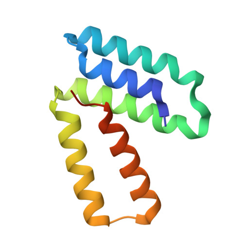
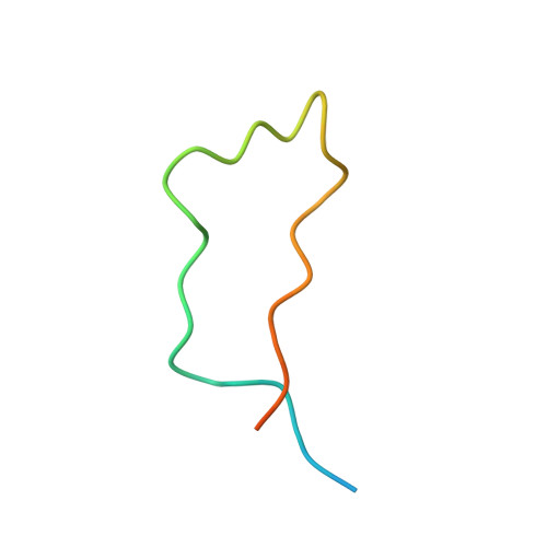
    The lectin chaperones calreticulin (CRT) and calnexin (CNX) contribute to the folding of glycoproteins in the ER by recruiting foldases such as the protein disulfide isomerase ERp57 and the peptidyl prolyl cis-trans isomerase CypB. Recently, CRT was shown to interact with the chaperone ERp29. Here, we show that ERp29 directly binds to the P domain of CNX. Crystal structures of the D domain of ERp29 in complex with the P domains from CRT and calmegin, a tissue-specific CNX homolog, reveal a commonality in the mechanism of binding whereby the tip of the P domain functions as a plurivalent adapter to bind a variety of folding factors. We show that mutation of a single residue, D348 in CNX, abrogates binding to ERp29 as well as ERp57 and CypB. The structural diversity of the accessory factors suggests that these chaperones became specialized for glycoprotein folding through convergent evolution of their P-domain binding sites.


  • Organizational Affiliation

    Department of Biochemistry, Groupe de recherche axé sur la Structure des protéines, McGill University, 3649 Promenade Sir William Osler, Montréal, QC H3G 0B1, Canada.


Macromolecules
Find similar proteins by:  (by identity cutoff)  |  3D Structure
Entity ID: 1
MoleculeChains Sequence LengthOrganismDetailsImage
Endoplasmic reticulum resident protein 29A [auth C],
B [auth A]
106Homo sapiensMutation(s): 0 
Gene Names: ERP29C12orf8ERP28
UniProt & NIH Common Fund Data Resources
Find proteins for P30040 (Homo sapiens)
Explore P30040 
Go to UniProtKB:  P30040
PHAROS:  P30040
GTEx:  ENSG00000089248 
Entity Groups  
Sequence Clusters30% Identity50% Identity70% Identity90% Identity95% Identity100% Identity
UniProt GroupP30040
Sequence Annotations
Expand
  • Reference Sequence
Find similar proteins by:  (by identity cutoff)  |  3D Structure
Entity ID: 2
MoleculeChains Sequence LengthOrganismDetailsImage
CalreticulinC [auth B],
D
40Homo sapiensMutation(s): 0 
Gene Names: CALRCRTC
UniProt & NIH Common Fund Data Resources
Find proteins for P27797 (Homo sapiens)
Explore P27797 
Go to UniProtKB:  P27797
PHAROS:  P27797
GTEx:  ENSG00000179218 
Entity Groups  
Sequence Clusters30% Identity50% Identity70% Identity90% Identity95% Identity100% Identity
UniProt GroupP27797
Sequence Annotations
Expand
  • Reference Sequence
Small Molecules
Ligands 1 Unique
IDChains Name / Formula / InChI Key2D Diagram3D Interactions
GOL
Query on GOL

Download Ideal Coordinates CCD File 
E [auth A]GLYCEROL
C3 H8 O3
PEDCQBHIVMGVHV-UHFFFAOYSA-N
Experimental Data & Validation

Experimental Data

  • Method: X-RAY DIFFRACTION
  • Resolution: 3.25 Å
  • R-Value Free: 0.262 
  • R-Value Work: 0.240 
  • R-Value Observed: 0.241 
  • Space Group: P 41 21 2
Unit Cell:
Length ( Å )Angle ( ˚ )
a = 68.573α = 90
b = 68.573β = 90
c = 167.778γ = 90
Software Package:
Software NamePurpose
PHENIXrefinement
HKL-2000data reduction
HKL-2000data scaling
PHASERphasing

Structure Validation

View Full Validation Report


Entry History & Funding Information

Deposition Data


Funding OrganizationLocationGrant Number
Natural Sciences and Engineering Research Council (NSERC, Canada)CanadaRGPIN 238873

Revision History  (Full details and data files)

  • Version 1.0: 2017-06-21
    Type: Initial release
  • Version 1.1: 2017-09-27
    Changes: Author supporting evidence
  • Version 1.2: 2018-10-03
    Changes: Data collection, Database references
  • Version 1.3: 2020-01-08
    Changes: Author supporting evidence
  • Version 1.4: 2023-10-04
    Changes: Data collection, Database references, Refinement description