5V2N

Crystal Structure of APO Human SETD8


Experimental Data Snapshot

  • Method: X-RAY DIFFRACTION
  • Resolution: 2.00 Å
  • R-Value Free: 0.250 
  • R-Value Work: 0.183 
  • R-Value Observed: 0.186 

wwPDB Validation   3D Report Full Report


This is version 1.2 of the entry. See complete history


Literature

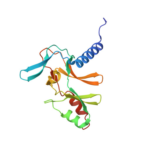
The dynamic conformational landscape of the protein methyltransferase SETD8.

Chen, S.Wiewiora, R.P.Meng, F.Babault, N.Ma, A.Yu, W.Qian, K.Hu, H.Zou, H.Wang, J.Fan, S.Blum, G.Pittella-Silva, F.Beauchamp, K.A.Tempel, W.Jiang, H.Chen, K.Skene, R.J.Zheng, Y.G.Brown, P.J.Jin, J.Luo, C.Chodera, J.D.Luo, M.

(2019) Elife 8

  • DOI: https://doi.org/10.7554/eLife.45403
  • Primary Citation of Related Structures:  
    5V2N, 6BOZ

  • PubMed Abstract: 

    Elucidating the conformational heterogeneity of proteins is essential for understanding protein function and developing exogenous ligands. With the rapid development of experimental and computational methods, it is of great interest to integrate these approaches to illuminate the conformational landscapes of target proteins. SETD8 is a protein lysine methyltransferase (PKMT), which functions in vivo via the methylation of histone and nonhistone targets. Utilizing covalent inhibitors and depleting native ligands to trap hidden conformational states, we obtained diverse X-ray structures of SETD8. These structures were used to seed distributed atomistic molecular dynamics simulations that generated a total of six milliseconds of trajectory data. Markov state models, built via an automated machine learning approach and corroborated experimentally, reveal how slow conformational motions and conformational states are relevant to catalysis. These findings provide molecular insight on enzymatic catalysis and allosteric mechanisms of a PKMT via its detailed conformational landscape.


  • Organizational Affiliation

    Tri-Institutional PhD Program in Chemical Biology, Memorial Sloan Kettering Cancer Center, New York, United States.


Macromolecules
Find similar proteins by:  (by identity cutoff)  |  3D Structure
Entity ID: 1
MoleculeChains Sequence LengthOrganismDetailsImage
N-lysine methyltransferase KMT5A165Homo sapiensMutation(s): 0 
Gene Names: KMT5APRSET7SET07SET8SETD8
EC: 2.1.1 (PDB Primary Data), 2.1.1.43 (PDB Primary Data)
UniProt & NIH Common Fund Data Resources
Find proteins for Q9NQR1 (Homo sapiens)
Explore Q9NQR1 
Go to UniProtKB:  Q9NQR1
PHAROS:  Q9NQR1
GTEx:  ENSG00000183955 
Entity Groups  
Sequence Clusters30% Identity50% Identity70% Identity90% Identity95% Identity100% Identity
UniProt GroupQ9NQR1
Sequence Annotations
Expand
  • Reference Sequence
Experimental Data & Validation

Experimental Data

  • Method: X-RAY DIFFRACTION
  • Resolution: 2.00 Å
  • R-Value Free: 0.250 
  • R-Value Work: 0.183 
  • R-Value Observed: 0.186 
  • Space Group: P 43 21 2
Unit Cell:
Length ( Å )Angle ( ˚ )
a = 60.62α = 90
b = 60.62β = 90
c = 80.708γ = 90
Software Package:
Software NamePurpose
HKL-2000data scaling
REFMACrefinement
PDB_EXTRACTdata extraction
HKL-2000data reduction
MOLREPphasing
HKLdata scaling

Structure Validation

View Full Validation Report


Entry History 

Deposition Data

  • Released Date: 2018-03-07 
  • Deposition Author(s): Skene, R.J.

Revision History  (Full details and data files)

  • Version 1.0: 2018-03-07
    Type: Initial release
  • Version 1.1: 2019-06-12
    Changes: Data collection, Database references
  • Version 1.2: 2024-03-06
    Changes: Data collection, Database references