5UCG

Structure of the PP2C Phosphatase Domain and a Fragment of the Regulatory Domain of the Cell Fate Determinant SpoIIE from Bacillus Subtilis


Experimental Data Snapshot

  • Method: X-RAY DIFFRACTION
  • Resolution: 3.91 Å
  • R-Value Free: 0.323 
  • R-Value Work: 0.276 
  • R-Value Observed: 0.279 

wwPDB Validation   3D Report Full Report


This is version 1.3 of the entry. See complete history


Literature

A widespread family of serine/threonine protein phosphatases shares a common regulatory switch with proteasomal proteases.

Bradshaw, N.Levdikov, V.M.Zimanyi, C.M.Gaudet, R.Wilkinson, A.J.Losick, R.

(2017) Elife 6

  • DOI: https://doi.org/10.7554/eLife.26111
  • Primary Citation of Related Structures:  
    5MQH, 5UCG

  • PubMed Abstract: 

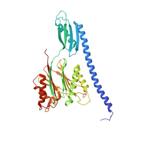
    PP2C phosphatases control biological processes including stress responses, development, and cell division in all kingdoms of life. Diverse regulatory domains adapt PP2C phosphatases to specific functions, but how these domains control phosphatase activity was unknown. We present structures representing active and inactive states of the PP2C phosphatase SpoIIE from Bacillus subtilis . Based on structural analyses and genetic and biochemical experiments, we identify an α-helical switch that shifts a carbonyl oxygen into the active site to coordinate a metal cofactor. Our analysis indicates that this switch is widely conserved among PP2C family members, serving as a platform to control phosphatase activity in response to diverse inputs. Remarkably, the switch is shared with proteasomal proteases, which we identify as evolutionary and structural relatives of PP2C phosphatases. Although these proteases use an unrelated catalytic mechanism, rotation of equivalent helices controls protease activity by movement of the equivalent carbonyl oxygen into the active site.


  • Organizational Affiliation

    Department of Molecular and Cellular Biology, Harvard University, Cambridge, United States.


Macromolecules
Find similar proteins by:  (by identity cutoff)  |  3D Structure
Entity ID: 1
MoleculeChains Sequence LengthOrganismDetailsImage
Stage II sporulation protein EA,
B [auth E],
C [auth B],
D [auth C],
E [auth D]
345Bacillus subtilis subsp. subtilis str. 168Mutation(s): 0 
Gene Names: spoIIEspoIIHBSU00640
EC: 3.1.3.16
UniProt
Find proteins for P37475 (Bacillus subtilis (strain 168))
Explore P37475 
Go to UniProtKB:  P37475
Entity Groups  
Sequence Clusters30% Identity50% Identity70% Identity90% Identity95% Identity100% Identity
UniProt GroupP37475
Sequence Annotations
Expand
  • Reference Sequence
Experimental Data & Validation

Experimental Data

Unit Cell:
Length ( Å )Angle ( ˚ )
a = 125.622α = 90
b = 125.622β = 90
c = 330.696γ = 90
Software Package:
Software NamePurpose
PHENIXrefinement
HKL-2000data scaling
PDB_EXTRACTdata extraction
HKL-2000data reduction
PHENIXphasing
DENZOdata reduction
SCALEPACKdata scaling

Structure Validation

View Full Validation Report


Entry History & Funding Information

Deposition Data


Funding OrganizationLocationGrant Number
National Institutes of Health/National Institute of General Medical Sciences (NIH/NIGMS)United StatesGM18568
Wellcome TrustUnited Kingdom082829
Damon Runyon Cancer Research FoundationUnited StatesDRG 2051-10
Jane Coffin Childs Memorial Fund for Medical ResearchUnited StatesJCC Fund

Revision History  (Full details and data files)

  • Version 1.0: 2017-05-31
    Type: Initial release
  • Version 1.1: 2017-09-20
    Changes: Author supporting evidence, Refinement description
  • Version 1.2: 2020-01-01
    Changes: Author supporting evidence
  • Version 1.3: 2023-10-04
    Changes: Data collection, Database references, Refinement description