5TXI

Crystal structure of wild-type S. aureus penicillin binding protein 4 (PBP4) in complex with ceftobiprole


Experimental Data Snapshot

  • Method: X-RAY DIFFRACTION
  • Resolution: 1.60 Å
  • R-Value Free: 0.202 
  • R-Value Work: 0.171 
  • R-Value Observed: 0.173 

wwPDB Validation   3D Report Full Report


Ligand Structure Quality Assessment 


This is version 1.3 of the entry. See complete history


Literature

Structural and kinetic analysis of penicillin-binding protein 4 (PBP4)-mediated antibiotic resistance inStaphylococcus aureus.

Alexander, J.A.N.Chatterjee, S.S.Hamilton, S.M.Eltis, L.D.Chambers, H.F.Strynadka, N.C.J.

(2018) J Biol Chem 

  • DOI: https://doi.org/10.1074/jbc.RA118.004952
  • Primary Citation of Related Structures:  
    5TW4, 5TW8, 5TX9, 5TXI, 5TY2, 5TY7, 6C39, 6C3K

  • PubMed Abstract: 

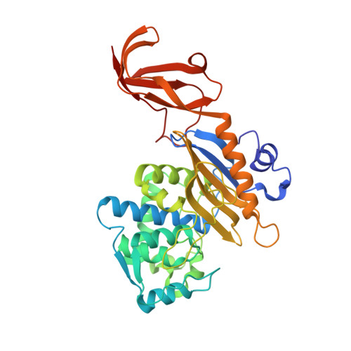
    Methicillin-resistant Staphylococcus aureus (MRSA) causes serious community-acquired and nosocomial infections worldwide. MRSA strains are resistant to a variety of antibiotics, including the classic penicillin and cephalosporin classes of β-lactams, making them intractable to treatment. Although β-lactam resistance in MRSA has been ascribed to the acquisition and activity of penicillin-binding protein 2a (PBP2a, encoded by mecA ), it has recently been observed that resistance can also be mediated by penicillin-binding protein 4 (PBP4). Previously, we have shown that broad-spectrum β-lactam resistance can arise following serial passaging of a mecA -negative COL strain of S. aureus, creating the CRB strain. This strain has two missense mutations in pbp4 and a mutation in the pbp4 promoter, both of which play an instrumental role in β-lactam resistance. To better understand PBP4's role in resistance, here we have characterized its kinetics and structure with clinically relevant β-lactam antibiotics. We present the first crystallographic PBP4 structures of apo and acyl-enzyme intermediate forms complexed with three late-generation β-lactam antibiotics: ceftobiprole, ceftaroline, and nafcillin. In parallel, we characterized the structural and kinetic effects of the PBP4 mutations present in the CRB strain. Localized within the transpeptidase active-site cleft, the two substitutions appear to have different effects depending on the drug. With ceftobiprole, the missense mutations impaired the K m value 150-fold, decreasing the proportion of inhibited PBP4. However, ceftaroline resistance appeared to be mediated by other factors, possibly including mutation of the pbp4 promoter. Our findings provide evidence that S. aureus CRB has at least two PBP4-mediated resistance mechanisms.


  • Organizational Affiliation

    From the Department of Biochemistry and Molecular Biology.


Macromolecules
Find similar proteins by:  (by identity cutoff)  |  3D Structure
Entity ID: 1
MoleculeChains Sequence LengthOrganismDetailsImage
Penicillin binding protein 4
A, B
359Staphylococcus aureusMutation(s): 0 
UniProt
Find proteins for Q2G2X6 (Staphylococcus aureus (strain NCTC 8325 / PS 47))
Explore Q2G2X6 
Go to UniProtKB:  Q2G2X6
Entity Groups  
Sequence Clusters30% Identity50% Identity70% Identity90% Identity95% Identity100% Identity
UniProt GroupQ2G2X6
Sequence Annotations
Expand
  • Reference Sequence
Small Molecules
Ligands 6 Unique
IDChains Name / Formula / InChI Key2D Diagram3D Interactions
RB6
Query on RB6

Download Ideal Coordinates CCD File 
C [auth A],
K [auth B]
(2R)-2-[(1R)-1-{[(2Z)-2-(5-amino-1,2,4-thiadiazol-3-yl)-2-(hydroxyimino)acetyl]amino}-2-oxoethyl]-5-({2-oxo-1-[(3R)-pyr rolidin-3-yl]-2,5-dihydro-1H-pyrrol-3-yl}methyl)-3,6-dihydro-2H-1,3-thiazine-4-carboxylic acid
C20 H24 N8 O6 S2
MYAXGJQBOYOEHQ-SWBIIUODSA-N
SO4
Query on SO4

Download Ideal Coordinates CCD File 
D [auth A],
L [auth B]
SULFATE ION
O4 S
QAOWNCQODCNURD-UHFFFAOYSA-L
GOL
Query on GOL

Download Ideal Coordinates CCD File 
Q [auth B]GLYCEROL
C3 H8 O3
PEDCQBHIVMGVHV-UHFFFAOYSA-N
ZN
Query on ZN

Download Ideal Coordinates CCD File 
E [auth A],
F [auth A],
G [auth A],
M [auth B]
ZINC ION
Zn
PTFCDOFLOPIGGS-UHFFFAOYSA-N
CL
Query on CL

Download Ideal Coordinates CCD File 
H [auth A],
N [auth B]
CHLORIDE ION
Cl
VEXZGXHMUGYJMC-UHFFFAOYSA-M
NA
Query on NA

Download Ideal Coordinates CCD File 
I [auth A],
J [auth A],
O [auth B],
P [auth B]
SODIUM ION
Na
FKNQFGJONOIPTF-UHFFFAOYSA-N
Experimental Data & Validation

Experimental Data

  • Method: X-RAY DIFFRACTION
  • Resolution: 1.60 Å
  • R-Value Free: 0.202 
  • R-Value Work: 0.171 
  • R-Value Observed: 0.173 
  • Space Group: C 1 2 1
Unit Cell:
Length ( Å )Angle ( ˚ )
a = 116.904α = 90
b = 92.293β = 100.01
c = 79.519γ = 90
Software Package:
Software NamePurpose
Aimlessdata scaling
PHASERphasing
PHENIXrefinement
PDB_EXTRACTdata extraction
XDSdata reduction

Structure Validation

View Full Validation Report


Ligand Structure Quality Assessment 


Entry History & Funding Information

Deposition Data


Funding OrganizationLocationGrant Number
Canadian Institutes of Health Research (CIHR)Canada--

Revision History  (Full details and data files)

  • Version 1.0: 2018-05-16
    Type: Initial release
  • Version 1.1: 2018-11-28
    Changes: Data collection, Database references
  • Version 1.2: 2020-01-08
    Changes: Author supporting evidence
  • Version 1.3: 2023-10-04
    Changes: Data collection, Database references, Derived calculations, Refinement description, Structure summary