5TOY

X-Ray Crystal Structure of Ruthenocene Conjugated Penicilloate and Penilloate Products in Complex with CTX-M-14 E166A Beta-Lactamase


Experimental Data Snapshot

  • Method: X-RAY DIFFRACTION
  • Resolution: 1.30 Å
  • R-Value Free: 0.163 
  • R-Value Work: 0.142 
  • R-Value Observed: 0.144 

wwPDB Validation   3D Report Full Report


Ligand Structure Quality Assessment 


This is version 2.1 of the entry. See complete history


Literature

Mechanisms of proton relay and product release by Class A beta-lactamase at ultrahigh resolution.

Lewandowski, E.M.Lethbridge, K.G.Sanishvili, R.Skiba, J.Kowalski, K.Chen, Y.

(2018) FEBS J 285: 87-100

  • DOI: https://doi.org/10.1111/febs.14315
  • Primary Citation of Related Structures:  
    5TOP, 5TOY, 5VLE

  • PubMed Abstract: 

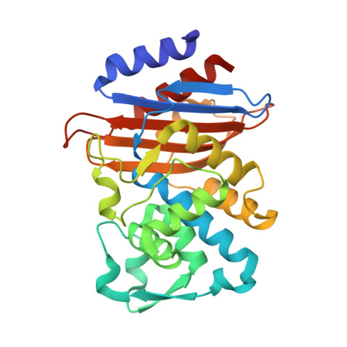
    The β-lactam antibiotics inhibit penicillin-binding proteins (PBPs) by forming a stable, covalent, acyl-enzyme complex. During the evolution from PBPs to Class A β-lactamases, the β-lactamases acquired Glu166 to activate a catalytic water and cleave the acyl-enzyme bond. Here we present three product complex crystal structures of CTX-M-14 Class A β-lactamase with a ruthenocene-conjugated penicillin-a 0.85 Å resolution structure of E166A mutant complexed with the penilloate product, a 1.30 Å resolution complex structure of the same mutant with the penicilloate product, and a 1.18 Å resolution complex structure of S70G mutant with a penicilloate product epimer-shedding light on the catalytic mechanisms and product inhibition of PBPs and Class A β-lactamases. The E166A-penilloate complex captured the hydrogen bonding network following the protonation of the leaving group and, for the first time, unambiguously show that the ring nitrogen donates a proton to Ser130, which in turn donates a proton to Lys73. These observations indicate that in the absence of Glu166, the equivalent lysine would be neutral in PBPs and therefore capable of serving as the general base to activate the catalytic serine. Together with previous results, this structure suggests a common proton relay network shared by Class A β-lactamases and PBPs, from the catalytic serine to the lysine, and ultimately to the ring nitrogen. Additionally, the E166A-penicilloate complex reveals previously unseen conformational changes of key catalytic residues during the release of the product, and is the first structure to capture the hydrolyzed product in the presence of an unmutated catalytic serine. Structural data are available in the PDB database under the accession numbers 5TOP, 5TOY, and 5VLE.


  • Organizational Affiliation

    Department of Molecular Medicine, University of South Florida College of Medicine, Tampa, FL, USA.


Macromolecules
Find similar proteins by:  (by identity cutoff)  |  3D Structure
Entity ID: 1
MoleculeChains Sequence LengthOrganismDetailsImage
Beta-lactamase
A, B
263Escherichia coliMutation(s): 1 
Gene Names: blaCTX-MCTX-M-14
EC: 3.5.2.6
UniProt
Find proteins for Q9L5C7 (Escherichia coli)
Explore Q9L5C7 
Go to UniProtKB:  Q9L5C7
Entity Groups  
Sequence Clusters30% Identity50% Identity70% Identity90% Identity95% Identity100% Identity
UniProt GroupQ9L5C7
Sequence Annotations
Expand
  • Reference Sequence
Oligosaccharides

Help

Entity ID: 2
MoleculeChains Length2D Diagram Glycosylation3D Interactions
beta-D-fructofuranose-(2-1)-alpha-D-glucopyranose
C, D, E
2N/A
Glycosylation Resources
GlyTouCan:  G05551OP
GlyCosmos:  G05551OP
Small Molecules
Ligands 5 Unique
IDChains Name / Formula / InChI Key2D Diagram3D Interactions
JSC
Query on JSC

Download Ideal Coordinates CCD File 
I [auth A],
N [auth B]
[(1,2,3,4,5-eta)-1-(4-{[carboxy(4-carboxy-5,5-dimethyl-1,3-thiazolidin-2-yl)methyl]amino}-4-oxobutanoyl)cyclopentadienyl][(1,2,3,4,5-eta)-cyclopentadienyl]ruthenium
C22 H17 N2 O6 Ru S
DONWCTWXNJLLEK-HTTVLNLHSA-N
JSD
Query on JSD

Download Ideal Coordinates CCD File 
J [auth A],
P [auth B]
[(1,2,3,4,5-eta)-1-(4-{[(4-carboxy-5,5-dimethyl-1,3-thiazolidin-2-yl)methyl]amino}-4-oxobutanoyl)cyclopentadienyl][(1,2,3,4,5-eta)-cyclopentadienyl]ruthenium
C21 H17 N2 O4 Ru S
JRWKHIBMJXULTB-BQFBZIMZSA-N
RU
Query on RU

Download Ideal Coordinates CCD File 
O [auth B]RUTHENIUM ION
Ru
BPEVHDGLPIIAGH-UHFFFAOYSA-N
PO4
Query on PO4

Download Ideal Coordinates CCD File 
G [auth A],
H [auth A],
L [auth B],
M [auth B]
PHOSPHATE ION
O4 P
NBIIXXVUZAFLBC-UHFFFAOYSA-K
K
Query on K

Download Ideal Coordinates CCD File 
F [auth A],
K [auth B]
POTASSIUM ION
K
NPYPAHLBTDXSSS-UHFFFAOYSA-N
Biologically Interesting Molecules (External Reference) 1 Unique
Experimental Data & Validation

Experimental Data

  • Method: X-RAY DIFFRACTION
  • Resolution: 1.30 Å
  • R-Value Free: 0.163 
  • R-Value Work: 0.142 
  • R-Value Observed: 0.144 
  • Space Group: P 1 21 1
Unit Cell:
Length ( Å )Angle ( ˚ )
a = 45.012α = 90
b = 106.787β = 101.76
c = 47.759γ = 90
Software Package:
Software NamePurpose
REFMACrefinement
HKL-2000data reduction
SCALEPACKdata scaling
PHASERphasing

Structure Validation

View Full Validation Report


Ligand Structure Quality Assessment 


Entry History & Funding Information

Deposition Data


Funding OrganizationLocationGrant Number
National Institutes of Health/National Institute Of Allergy and Infectious Diseases (NIH/NIAID)United StatesAI03158
Polish National Science CentrePolandDEC-2013/11/B/ST5/00997

Revision History  (Full details and data files)

  • Version 1.0: 2017-11-08
    Type: Initial release
  • Version 1.1: 2017-11-15
    Changes: Database references
  • Version 1.2: 2018-01-17
    Changes: Database references
  • Version 1.3: 2019-12-11
    Changes: Author supporting evidence
  • Version 2.0: 2020-07-29
    Type: Remediation
    Reason: Carbohydrate remediation
    Changes: Atomic model, Data collection, Derived calculations, Non-polymer description, Structure summary
  • Version 2.1: 2023-10-04
    Changes: Data collection, Database references, Refinement description, Structure summary