5T13

Structure of the Cyanuric Acid Hydrolase TrzD Reveals Product Exit Channel


Experimental Data Snapshot

  • Method: X-RAY DIFFRACTION
  • Resolution: 2.19 Å
  • R-Value Free: 0.200 
  • R-Value Work: 0.152 
  • R-Value Observed: 0.154 

wwPDB Validation   3D Report Full Report


This is version 1.1 of the entry. See complete history


Literature

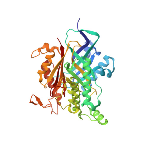
Structure of the Cyanuric Acid Hydrolase TrzD Reveals Product Exit Channel.

Bera, A.K.Aukema, K.G.Elias, M.Wackett, L.P.

(2017) Sci Rep 7: 45277-45277

  • DOI: https://doi.org/10.1038/srep45277
  • Primary Citation of Related Structures:  
    5T13

  • PubMed Abstract: 

    Cyanuric acid hydrolases are of industrial importance because of their use in aquatic recreational facilities to remove cyanuric acid, a stabilizer for the chlorine. Degradation of excess cyanuric acid is necessary to maintain chlorine disinfection in the waters. Cyanuric acid hydrolase opens the cyanuric acid ring hydrolytically and subsequent decarboxylation produces carbon dioxide and biuret. In the present study, we report the X-ray structure of TrzD, a cyanuric acid hydrolase from Acidovorax citrulli. The crystal structure at 2.19 Å resolution shows a large displacement of the catalytic lysine (Lys163) in domain 2 away from the active site core, whereas the two other active site lysines from the two other domains are not able to move. The lysine displacement is proposed here to open up a channel for product release. Consistent with that, the structure also showed two molecules of the co-product, carbon dioxide, one in the active site and another trapped in the proposed exit channel. Previous data indicated that the domain 2 lysine residue plays a role in activating an adjacent serine residue carrying out nucleophilic attack, opening the cyanuric acid ring, and the mobile lysine guides products through the exit channel.


  • Organizational Affiliation

    BioTechnology Institute, University of Minnesota, St. Paul, MN 55108, USA.


Macromolecules
Find similar proteins by:  (by identity cutoff)  |  3D Structure
Entity ID: 1
MoleculeChains Sequence LengthOrganismDetailsImage
Cyanuric acid amidohydrolase376Enterobacter cloacaeMutation(s): 0 
Gene Names: trzD
EC: 3.5.2.15
UniProt
Find proteins for P0A3V4 (Enterobacter cloacae)
Explore P0A3V4 
Go to UniProtKB:  P0A3V4
Entity Groups  
Sequence Clusters30% Identity50% Identity70% Identity90% Identity95% Identity100% Identity
UniProt GroupP0A3V4
Sequence Annotations
Expand
  • Reference Sequence
Experimental Data & Validation

Experimental Data

  • Method: X-RAY DIFFRACTION
  • Resolution: 2.19 Å
  • R-Value Free: 0.200 
  • R-Value Work: 0.152 
  • R-Value Observed: 0.154 
  • Space Group: I 2 2 2
Unit Cell:
Length ( Å )Angle ( ˚ )
a = 67.591α = 90
b = 90.686β = 90
c = 120.075γ = 90
Software Package:
Software NamePurpose
PHENIXrefinement
HKL-2000data reduction
HKL-2000data scaling
MOLREPphasing

Structure Validation

View Full Validation Report


Entry History & Funding Information

Deposition Data


Funding OrganizationLocationGrant Number
MnDRIVE Transdisciplinary Research grantUnited States--

Revision History  (Full details and data files)

  • Version 1.0: 2017-04-12
    Type: Initial release
  • Version 1.1: 2023-10-04
    Changes: Data collection, Database references, Refinement description