5PTI

STRUCTURE OF BOVINE PANCREATIC TRYPSIN INHIBITOR. RESULTS OF JOINT NEUTRON AND X-RAY REFINEMENT OF CRYSTAL FORM II


Experimental Data Snapshot

  • Method: X-RAY DIFFRACTION
  • Resolution: 1.00 Å

    wwPDB Validation   3D Report Full Report


    This is version 1.4 of the entry. See complete history


    Literature

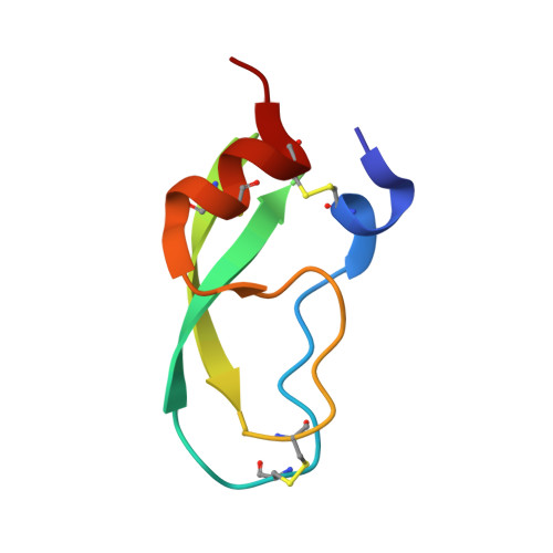
    Structure of bovine pancreatic trypsin inhibitor. Results of joint neutron and X-ray refinement of crystal form II

    Wlodawer, A.Walter, J.Huber, R.Sjolin, L.

    (1984) J Mol Biol 180: 301-329

    • DOI: https://doi.org/10.1016/s0022-2836(84)80006-6
    • Primary Citation of Related Structures:  
      5PTI

    • PubMed Abstract: 

      The structure of form II crystals of bovine pancreatic trypsin inhibitor has been investigated by joint refinement of X-ray and neutron data. Crystallographic R factors for the final model were 0.200 for the X-ray data extending to 1 A resolution and 0.197 for the 1.8 A neutron data. This model was strongly restrained, with 0.020 A root-mean-square (r.m.s.) departure of bond lengths from their ideal values and 0.019 A r.m.s. departure of planar groups from planarity. The resulting structure was very similar to that of crystal form I (r.m.s. deviation for main chain atoms was 0.40 A); nevertheless larger deviations were observed in particular regions of the chain. Twenty out of 63 ordered water molecules occupy similar positions (deviation less than 1 A) in both models. Eleven amide hydrogens were found to be protected from exchange after three months of soaking the crystals in deuterated mother liquor at pH 8.2. Their locations were in excellent agreement with the results obtained by two-dimensional nuclear magnetic resonance, but the rates of exchange are much lower in the crystalline state.


    Macromolecules
    Find similar proteins by:  (by identity cutoff)  |  3D Structure
    Entity ID: 1
    MoleculeChains Sequence LengthOrganismDetailsImage
    TRYPSIN INHIBITOR58Bos taurusMutation(s): 0 
    UniProt
    Find proteins for P00974 (Bos taurus)
    Explore P00974 
    Go to UniProtKB:  P00974
    Entity Groups  
    Sequence Clusters30% Identity50% Identity70% Identity90% Identity95% Identity100% Identity
    UniProt GroupP00974
    Sequence Annotations
    Expand
    • Reference Sequence
    Small Molecules
    Ligands 2 Unique
    IDChains Name / Formula / InChI Key2D Diagram3D Interactions
    PO4
    Query on PO4

    Download Ideal Coordinates CCD File 
    B [auth A]PHOSPHATE ION
    O4 P
    NBIIXXVUZAFLBC-UHFFFAOYSA-K
    UNX
    Query on UNX

    Download Ideal Coordinates CCD File 
    C [auth A]UNKNOWN ATOM OR ION
    X
    Experimental Data & Validation

    Experimental Data

    • Method: X-RAY DIFFRACTION
    • Resolution: 1.00 Å
    • Space Group: P 21 21 21
    Unit Cell:
    Length ( Å )Angle ( ˚ )
    a = 74.1α = 90
    b = 23.4β = 90
    c = 28.9γ = 90

    Structure Validation

    View Full Validation Report


    Entry History 

    Deposition Data

    Revision History  (Full details and data files)

    • Version 1.0: 1984-10-29
      Type: Initial release
    • Version 1.1: 2008-03-27
      Changes: Version format compliance
    • Version 1.2: 2011-07-13
      Changes: Version format compliance
    • Version 1.3: 2011-07-27
      Changes: Atomic model, Data collection
    • Version 1.4: 2018-10-24
      Changes: Data collection, Other, Structure summary