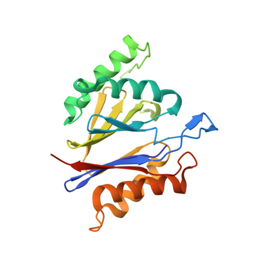
Structural characterization of the bacterial proteasome homolog BPH reveals a tetradecameric double-ring complex with unique inner cavity properties.
Fuchs, A.C.D., Maldoner, L., Hipp, K., Hartmann, M.D., Martin, J.(2018) J Biol Chem 293: 920-930
- PubMed: 29183996
- DOI: https://doi.org/10.1074/jbc.M117.815258
- Primary Citation of Related Structures:
5OVS, 5OVT, 5OVU - PubMed Abstract:
Eukaryotic and archaeal proteasomes are paradigms for self-compartmentalizing proteases. To a large extent, their function requires interplay with hexameric ATPases associated with diverse cellular activities (AAA+) that act as substrate unfoldases. Bacteria have various types of self-compartmentalizing proteases; in addition to the proteasome itself, these include the proteasome homolog HslV, which functions together with the AAA+ HslU; the ClpP protease with its partner AAA+ ClpX; and Anbu, a recently characterized ancestral proteasome variant. Previous bioinformatic analysis has revealed a novel bacterial member of the proteasome family Betaproteobacteria proteasome homolog (BPH). Using cluster analysis, we here affirmed that BPH evolutionarily descends from HslV. Crystal structures of the Thiobacillus denitrificans and Cupriavidus metallidurans BPHs disclosed a homo-oligomeric double-ring architecture in which the active sites face the interior of the cylinder. Using small-angle X-ray scattering (SAXS) and electron microscopy averaging, we found that BPH forms tetradecamers in solution, unlike the dodecamers seen in HslV. Although the highly acidic inner surface of BPH was in striking contrast to the cavity characteristics of the proteasome and HslV, a classical proteasomal reaction mechanism could be inferred from the covalent binding of the proteasome-specific inhibitor epoxomicin to BPH. A ligand-bound structure implied that the elongated BPH inner pore loop may be involved in substrate recognition. The apparent lack of a partner unfoldase and other unique features, such as Ser replacing Thr as the catalytic residue in certain BPH subfamilies, suggest a proteolytic function for BPH distinct from those of known bacterial self-compartmentalizing proteases.
Organizational Affiliation:
From the Department of Protein Evolution and.