5NJL

Cwp2 from Clostridium difficile


Experimental Data Snapshot

  • Method: X-RAY DIFFRACTION
  • Resolution: 1.90 Å
  • R-Value Free: 0.245 
  • R-Value Work: 0.215 
  • R-Value Observed: 0.216 

wwPDB Validation   3D Report Full Report


This is version 1.5 of the entry. See complete history


Literature

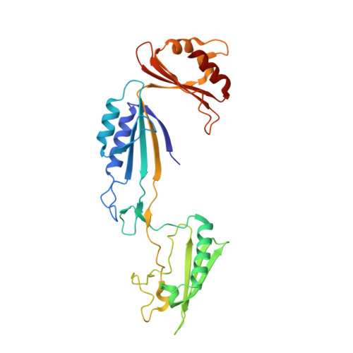
Cwp2 from Clostridium difficile exhibits an extended three domain fold and cell adhesion in vitro.

Bradshaw, W.J.Kirby, J.M.Roberts, A.K.Shone, C.C.Acharya, K.R.

(2017) FEBS J 284: 2886-2898

  • DOI: https://doi.org/10.1111/febs.14157
  • Primary Citation of Related Structures:  
    5NJL

  • PubMed Abstract: 

    Colonization of the gut by Clostridium difficile requires the adhesion of the bacterium to host cells. A range of cell surface located factors have been linked to adhesion including the S-layer protein LMW SLP and the related protein Cwp66. As well as these proteins, the S-layer of C. difficile may contain many others. One such protein is Cwp2. Here, we demonstrate the production of a C. difficile strain 630 cwp2 knockout mutant and assess the effect on the bacterium. The mutant results in increased TcdA (toxin A) release and impaired cellular adherence in vitro. We also present the extended three domain structure of the 'functional' region of Cwp2, consisting of residues 29-318 at 1.9 Å, which is compared to that of LMW SLP and Cwp8. The adhesive properties of Cwp2 and LMW SLP, which are likely to be shared by Cwp8, are predicted to be mediated by the variable loop regions in domain 2.


  • Organizational Affiliation

    Department of Biology and Biochemistry, University of Bath, UK.


Macromolecules
Find similar proteins by:  (by identity cutoff)  |  3D Structure
Entity ID: 1
MoleculeChains Sequence LengthOrganismDetailsImage
Cell surface protein (Putative S-layer protein)295Clostridioides difficile R20291Mutation(s): 0 
Gene Names: CDR20291_2680
Entity Groups  
Sequence Clusters30% Identity50% Identity70% Identity90% Identity95% Identity100% Identity
Sequence Annotations
Expand
  • Reference Sequence
Small Molecules
Ligands 1 Unique
IDChains Name / Formula / InChI Key2D Diagram3D Interactions
SO4
Query on SO4

Download Ideal Coordinates CCD File 
B [auth A]
C [auth A]
D [auth A]
E [auth A]
F [auth A]
B [auth A],
C [auth A],
D [auth A],
E [auth A],
F [auth A],
G [auth A],
H [auth A],
I [auth A],
J [auth A],
K [auth A],
L [auth A],
M [auth A],
N [auth A],
O [auth A],
P [auth A],
Q [auth A],
R [auth A],
S [auth A],
T [auth A],
U [auth A]
SULFATE ION
O4 S
QAOWNCQODCNURD-UHFFFAOYSA-L
Experimental Data & Validation

Experimental Data

  • Method: X-RAY DIFFRACTION
  • Resolution: 1.90 Å
  • R-Value Free: 0.245 
  • R-Value Work: 0.215 
  • R-Value Observed: 0.216 
  • Space Group: P 63 2 2
Unit Cell:
Length ( Å )Angle ( ˚ )
a = 134.828α = 90
b = 134.828β = 90
c = 102.835γ = 120
Software Package:
Software NamePurpose
REFMACrefinement
DIALSdata reduction
Aimlessdata scaling
PHASERphasing

Structure Validation

View Full Validation Report


Entry History & Funding Information

Deposition Data


Funding OrganizationLocationGrant Number
Medical Research Council (United Kingdom)United KingdomMK/K027123/1

Revision History  (Full details and data files)

  • Version 1.0: 2017-07-05
    Type: Initial release
  • Version 1.1: 2017-07-19
    Changes: Database references
  • Version 1.2: 2017-09-13
    Changes: Author supporting evidence
  • Version 1.3: 2017-09-20
    Changes: Database references
  • Version 1.4: 2018-01-31
    Changes: Structure summary
  • Version 1.5: 2024-01-17
    Changes: Data collection, Database references, Refinement description